This site uses cookies to improve your experience. To help us insure we adhere to various privacy regulations, please select your country/region of residence. If you do not select a country, we will assume you are from the United States. Select your Cookie Settings or view our Privacy Policy and Terms of Use.
Cookie Settings
Cookies and similar technologies are used on this website for proper function of the website, for tracking performance analytics and for marketing purposes. We and some of our third-party providers may use cookie data for various purposes. Please review the cookie settings below and choose your preference.
Used for the proper function of the website
Used for monitoring website traffic and interactions
Cookie Settings
Cookies and similar technologies are used on this website for proper function of the website, for tracking performance analytics and for marketing purposes. We and some of our third-party providers may use cookie data for various purposes. Please review the cookie settings below and choose your preference.
Strictly Necessary: Used for the proper function of the website
Performance/Analytics: Used for monitoring website traffic and interactions
Nearly 72,000 chest x-rays had been randomized as of November 25 (the study is open through December 31), with the two primary outcomes of the trial being time to diagnosis of lung cancer and time from chest x-ray to CT by prioritizing abnormals. UCLH has produced 9,217 chest x-rays from 8,072 patients.
In a study described as a “competition between radiologists,” participants tasked with identifying abnormal findings on chest x-rays performed better with AI assistance than without AI assistance – though not by much and not in all cases, according to a group in Nanjing, Jiangsu, China.
Reading Time: 10 minutes read By Henry Williams, Carestream Area Vice President, Sales Western Nowadays, with hospital budgetary restrictions at the forefront of the purchasing decision making process, it seems like the X-Ray market, like everything else, is not immune to the current state of the economy. But is that really the case?
12, 2025 Konica Minolta Healthcare Americas, has published a case study by clinicians in the pulmonary and radiology departments at ASST Fatebenefratelli Sacco (Milan, Italy) demonstrating the use of Dynamic Digital Radiography (DDR) to help definitively diagnose diaphragm dysfunction. tim.hodson Fri, 02/14/2025 - 15:14 Feb.12,
Diagnostic imaging tests are tools used by physicians to diagnose a range of medical conditions. For X-rays, it usually takes less than 10 minutes. Why Diagnostic Imaging Methods Are Important Medical imaging is used to diagnose, monitor, and treat medical problems. It can take up to 90 minutes, max.
Common diagnostic tests for pulmonary disorders include chest x-rays and pulmonary function tests (PFTs). a) Raw example of a dynamic digital radiograph. (b) The digital technology limits radiation exposure to patients compared with standard chest x-rays, they wrote.
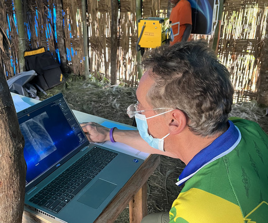
milla1cf Mon, 04/01/2024 - 11:44 April 1, 2024 — MinXray , a leading manufacturer of imaging systems for medical and veterinary use, recently sent its Impact Wireless X-ray system with a group of researchers and medical personnel to the YUS Conservation Area in Papua New Guinea.
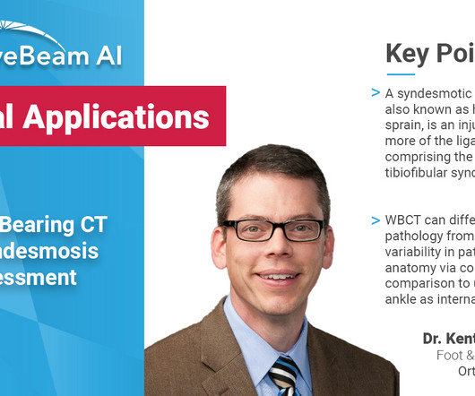
Key Points: Weight Bearing (WB) radiographs are limited by 1) their 2D nature and 2) knee positioning. X-Ray (XR) beam angle differs by person and can be unreliable. Optimal knee positioning and X-Ray beam angle differs by person and can be unreliable with WB radiographs. Segal et al.
Morphological markers on MRI help diagnose autism. Commercially Available Chest Radiograph AI Tools for Detecting Airspace Disease, Pneumothorax, and Pleural Effusion. Generative Artificial Intelligence for Chest Radiograph Interpretation in the Emergency Department. Cognitive Motor Dissociation in Disorders of Consciousness.
Dubai-based Zscale Labs is launching Neuromorphic AI, an AI software application for multilabel classification of chest radiographs. Neuromorphic AI leverages the firm's hyperdimensional computing and deep-learning technologies to assist radiologists and healthcare providers in diagnosing multiple chest conditions from x-ray images.
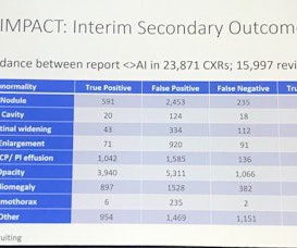
a key global supplier of portable, compact digital imaging equipment, has earned a Guinness World Record for the “ highest altitude operating an X-ray machine ” while on an expedition with both MinXray and artificial intelligence company Qure.ai. to demonstrate portable digital radiography and its effectiveness at high altitudes.
It uses AI to analyze CT scans, X-rays and pathology slides, supporting clinicians in detecting and diagnosing medical conditions faster and more accurately. Effect of a comprehensive deep-learning model on the accuracy of chest x-ray interpretation by radiologists: a retrospective, multireader multicase study.
Teleradiology-India Introduction: “X-ray Visionaries” takes you on a compelling journey to unveil the expertise of radiographers and technologists, the unsung heroes of X-ray technology. The significance of their expertise in ensuring accurate diagnoses and patient well-being.
Foot X-rays are the most common method podiatrists use to diagnose injuries and other conditions. If a patient is unable to take four steps on their foot without support, that’s a sure sign an X-ray, also called a radiograph, is needed.
Introduction: Delve into the illuminating world of X-rays and their pivotal role in diagnosing and monitoring Inflammatory Bowel Disease (IBD). This exploration sheds light on the radiological insights that X-ray imaging provides, offering valuable information for the comprehensive understanding and management of IBD. **1.
A team led by Avneesh Chhabra, MD, of the University of Texas Southwestern Medical Center in Dallas, found that the system's overall accuracy was higher than final diagnoses rendered without using it, at 65% compared with 55%. The study results were published August 27 in Radiology.
A team led by Alison Chang of Northwestern University Feinberg School of Medicine in Chicago reported that baseline MR imaging showed structural joint changes that did not appear on x-ray imaging -- the go-to modality for detecting the condition -- but the modality's ability to predict whether the disease would progress wasn't strong.
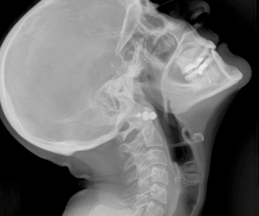
Lumbosacral spine X-rays, also called lumbar spine X-rays, are a radiographic imaging technique that uses low doses of electromagnetic radiation to view the internal anatomy of the lower spine, called the lumbosacral region.
Introduction: Dental X-ray technologists, also known as dental radiographers, are the unsung heroes behind the scenes of every successful dental diagnosis and treatment. In this blog, we’ll take you on a journey inside the jaw and explore a day in the life of a dental X-ray technologist.
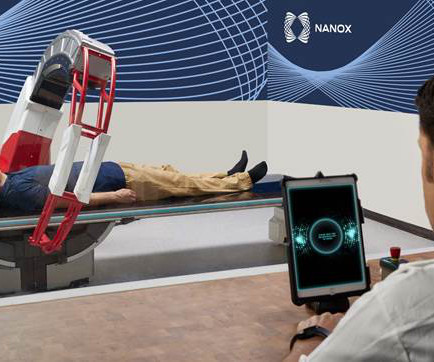
milla1cf Mon, 05/01/2023 - 17:39 May 1, 2023 — Nano-X Imaging Ltd. , Nanox.ARC is a stationary X-ray system intended to produce tomographic images of the human musculoskeletal system adjunctive to conventional radiography on adult patients. While access to medical imaging is relatively high in the U.S.
“Knowing that the industry continues to contend with staffing shortages and reduced budgets, Carestream is highlighting our advanced smart features that help radiographers execute quality exams in less time with less fatigue, leaving more time for patient interaction.
Closeup of X-ray photography of human brain Introduction: In the world of modern medicine, there exists a fascinating blend of art and science, where the careful use of technology and technique converges to reveal the hidden truths within the human body. Radiographic film, once the primary medium, has given way to digital sensors.
A patient’s specific needs and concerns are assessed, and a personalized radiographic plan is developed, taking into account factors like age, health conditions, and pregnancy. These methods provide comprehensive views of the oral cavity, enabling precise diagnoses and treatment planning.
milla1cf Sun, 10/01/2023 - 17:03 September 29, 2023 — Qure.ai , a leading global innovator in radiology AI solutions, has announced FDA clearance for measuring the cardiothoracic ratio (CTR) utilizing its artificial intelligence-enabled chest X-ray solution, qXR-CTR.
milla1cf Thu, 06/06/2024 - 21:32 June 6, 2024 — In a landmark study, the latest in technology innovation by Konica Minolta Healthcare was used to develop a machine-learning-based analysis of X-ray imaging that automatically quantifies lung function data. Chest radiography is typically acquired in the evaluation of pulmonary disorders.
In the third blog of her series on AI and the radiographer, Shamie Kumar explores the impact on the radiographer when AI is integrated within an imaging modality. The question to explore in this blog is when AI is integrated within an imaging modality itself and how that may impact a radiographer.
X-Ray Cone Beam CT WBCT can be used to determine places of instability throughout the foot and ankle, which may help to guide surgical reconstruction. Non-WB radiographs could not determine instability or severity of midfoot collapse; surgical intervention postponed. The patient was diagnosed with Charcot arthropathy.
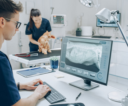
Veterinary technicians play a pivotal role in the radiographic process within veterinary practices. Their expertise in handling animals, positioning them correctly, and ensuring optimal image quality is essential for accurate diagnoses. Double-Check Alignment : Confirm that the area of interest is correctly aligned with the x-ray beam.
A weight bearing CT scan can: Provide increased sensitivity and specificity over radiographs. Diagnosis If WB X-rays are indeterminate after clinical exam, order a bilateral WBCT. X-rays were non-diagnostic. Utility of WBCT to Diagnose Syndesmotic Instability in Patients With Weber B Lateral Malleolar Fractures.
Many cases are diagnosed when it's too late, and a fracture has already occurred. Researchers at Naitive Technologies have invented a technique powered by artificial intelligence that extracts new, clinically valuable information from these routine X-rays without requiring additional equipment or physician time.
Through the use of iodine-based x-ray contrast agents, CEM can allow for better visualization of abnormalities in breast tissue that may not be visible with standard mammography.3 million women diagnosed with breast cancer globally, according to the World Health Organization (WHO).4 RadioGraphics 2019 39:7, 1907-1920.
A radiographer is a medical professional who performs the scanning on patients. Digital X-rays use less radiation and are employed for the same purposes. Digital X-rays use less radiation and are employed for the same purposes.
It all started when Wilhelm Conrad Röntgen discovered X-rays in 1895. After working for weeks in his lab experimenting on the production of ‘strange rays’, which he referred to as ‘X’, he asked his wife Anna Bertha to lend ‘a hand’, the left one to be precise, which he used to produce the first X-ray image.
Significance of Chest Radiography in Respiratory Health: Establish the pivotal role of chest radiography in diagnosing and monitoring respiratory conditions, with a focus on pneumonia. Discuss the ability of deep learning algorithms to autonomously learn and analyze complex patterns in radiographic images.
Occult pneumothoraces are those not suspected clinically or not evident on plain radiographs but later identified on computerized tomography (CT) imaging. Inclusion Criteria Patients of any age diagnosed with a blunt or penetrating traumatic occult pneumothorax on thoracoabdominal CT scan receiving mechanical ventilation.
According to a paper published in RadioGraphics , deep learning has enhanced the accuracy and efficiency of medical image interpretation, aiding radiologists in diagnosing conditions such as breast cancer, brain tumors, interstitial lung disease and intracranial hemorrhages.
A growing number of medical organisations link to the DenseBreast-info.org website, including the EFRS (European Federation of Radiographer Societies) and the Society of Radiographers. Figure 4 (b) The website includes breast screening guidelines in Europe. A comparative analysis table summarises the guidelines in each country.
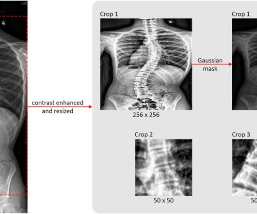
The deformity is usually diagnosed during puberty. Thus, the researchers aimed to develop an AI neural network that can automatically predict the probability of in-brace curve progression of AIS patients at their first visit to orthopedic clinics by using both routinely collected 1D clinical data and 2D full-spine x-ray images.
Our] results demonstrate [residents'] difficulties in accurately diagnosing these fractures, with observational errors being the primary issue across all cases and training levels," the group wrote. The team reported the following: 321 residents evaluated sacral ala fracture x-rays and received an average score of 1.29
What if X-ray imaging, the most prevalent and accessible imaging modality in the world, could provide the information needed for diagnosis? This unique study will pair X-rays of consented patients with their high-resolution CTs (HRCTs) as well as weekly forced vital capacity (FVC) readings collected via an app for home spirometry.
Conventional radiographs and MRIs have been the standard, but they come with limitations when it comes to understanding the complex, three-dimensional structure of the human body. As highlighted in the video, traditional X-rays provide only a two-dimensional view of three-dimensional structures. Why Choose WBCT?
Key Points: Imaging modalities such as plain radiographs (X-Ray), computed tomography (CT), and magnetic resonance imaging (MRI), dont have the diagnostic accuracy needed to detect syndesmotic widening or subtle instability. Method Nineteen matched pairs of through-the-knee cadaveric specimens (38 legs) were used in this study.
We organize all of the trending information in your field so you don't have to. Join 5,000 users and stay up to date on the latest articles your peers are reading.
You know about us, now we want to get to know you!
Let's personalize your content
Let's get even more personalized
We recognize your account from another site in our network, please click 'Send Email' below to continue with verifying your account and setting a password.


Let's personalize your content