This site uses cookies to improve your experience. To help us insure we adhere to various privacy regulations, please select your country/region of residence. If you do not select a country, we will assume you are from the United States. Select your Cookie Settings or view our Privacy Policy and Terms of Use.
Cookie Settings
Cookies and similar technologies are used on this website for proper function of the website, for tracking performance analytics and for marketing purposes. We and some of our third-party providers may use cookie data for various purposes. Please review the cookie settings below and choose your preference.
Used for the proper function of the website
Used for monitoring website traffic and interactions
Cookie Settings
Cookies and similar technologies are used on this website for proper function of the website, for tracking performance analytics and for marketing purposes. We and some of our third-party providers may use cookie data for various purposes. Please review the cookie settings below and choose your preference.
Strictly Necessary: Used for the proper function of the website
Performance/Analytics: Used for monitoring website traffic and interactions
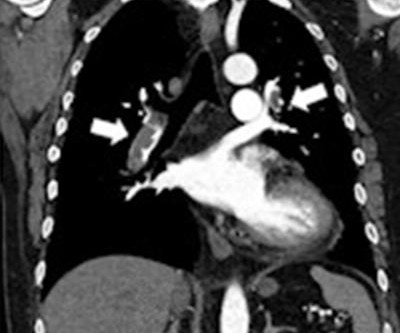
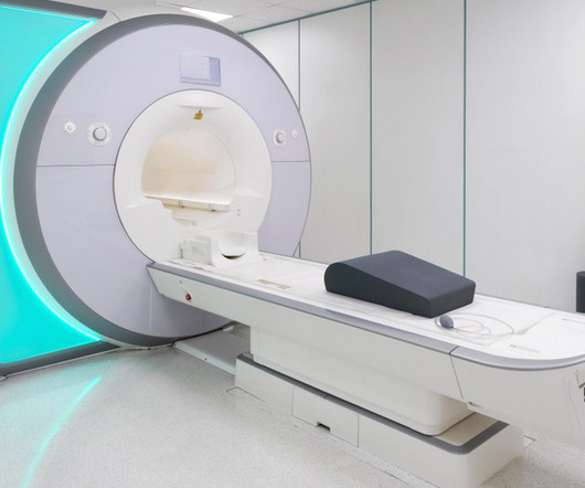
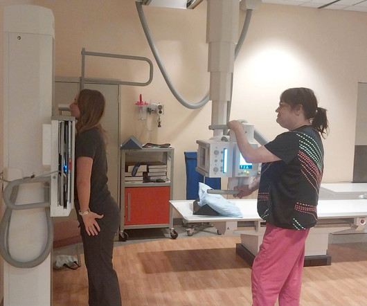
12, 2025 Konica Minolta Healthcare Americas, has published a case study by clinicians in the pulmonary and radiology departments at ASST Fatebenefratelli Sacco (Milan, Italy) demonstrating the use of Dynamic Digital Radiography (DDR) to help definitively diagnose diaphragm dysfunction. tim.hodson Fri, 02/14/2025 - 15:14 Feb.12,
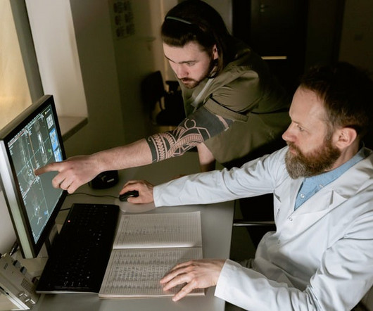
Researchers at the Icahn School of Medicine at Mount Sinai ("Icahn Mount Sinai") used Dynamic Digital Radiography (DDR) data, an X-ray imaging technology developed by Konica Minolta, to create their AI-powered technique that analyzes lung function. Chest radiography is typically acquired in the evaluation of pulmonary disorders.
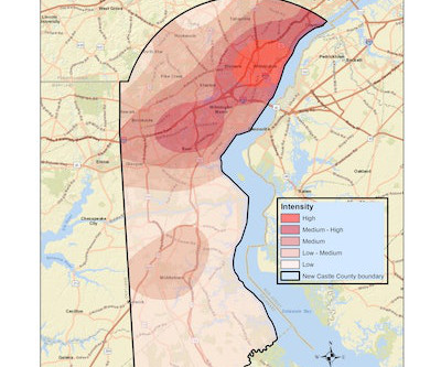
While Black and white women are diagnosed with breast cancer at similar rates, previous research suggests that Black women have a 40% higher mortality rate. in the incidence of triple-negative breast cancer and advanced breast cancer diagnosed at a younger age, ChristianaCare researchers highlighted. Delaware leads the U.S.
Imaging tests are a first-line tool for determining diagnoses, but the researchers underscored that health practitioners “often perform poorly” at estimating probabilities of disease before and after imaging exams are performed.
Chest dynamic digital radiography (DDR) may have received a boost toward clinical use in patients with lung disorders, with researchers developing AI to perform time-consuming analysis involved in the technology, according to researchers in New York City. Ultimately, DDR has unique advantages over traditional studies, the authors noted.
. | W3-SSMK08 | Room 5E450A In this session on musculoskeletal imaging, a deep-learning AI algorithm will be presented for measuring shoulder kinematics using dynamic digital radiography (DDR) images. Deep-learning model can spot patients at high risk of COPD Thursday, November 30 | 8:20 a.m.-8:30 8:30 a.m. | 9:20 a.m. | 1:50 p.m. |
As technology continues to advance, physicians have a multitude of methods to diagnose diseases. Radiography is a broad field that uses imaging techniques to view the skin. Doctors use diagnostic radiography to non-invasively diagnose injuries and diseases inside the body. Enter diagnostic radiology.
Understanding the distinction between radiography and sonography is vital in the increasingly specialized and multifaceted world of medical imaging. Radiography uses ionizing radiation to capture detailed internal images, which helps diagnose various conditions.
It uses AI to analyze CT scans, X-rays and pathology slides, supporting clinicians in detecting and diagnosing medical conditions faster and more accurately. aims to address the global shortage of 1.5 million skilled clinicians and the surge in demand for diagnosis impacting both developed and under-developed healthcare systems.
X-ray radiography is a noninvasive diagnostic method that uses X-rays—electromagnetic radiation—to produce images of the body's internal structures. Essential in medical fields for diagnosing injuries and diseases and monitoring treatment progress, X-ray radiography is a critical tool in patient care and medical decision-making.
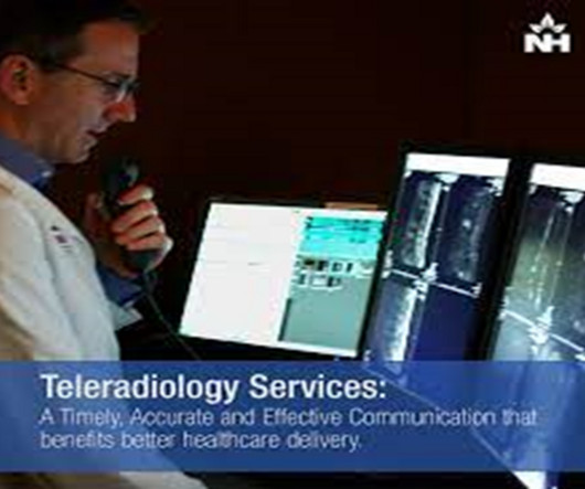
Teleradiology Introduction: Dental radiography plays a pivotal role in modern dentistry, enabling practitioners to diagnose and treat various oral health conditions. However, the field of dental radiography has undergone significant advancements, along with its own set of challenges and triumphs.
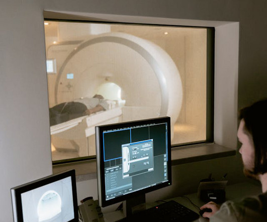
MRI-Scan-Teleradiology Introduction: Dental radiography is an essential component of modern dentistry, offering valuable insights into oral health and guiding treatments with precision. Patient-Centered Approach: Dental radiography starts with the patient. This quality control is vital for accurate diagnoses.
Researchers have defined a set of ultrasound parameters that quantitatively evaluate various physical lung characteristics for a more accurate assessment.
Radiography is at the forefront of modern medical diagnostics, a rapidly evolving field with innovative technological advancements. Its significance in healthcare is unparalleled, serving as a crucial tool in diagnosing and managing many health conditions.
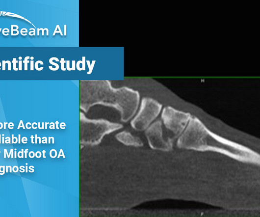
They found that midfoot OA often went completely undetected on weight bearing X-Ray and that WBCT helped diagnose and grade earlier, and more reliably, the onset of midfoot OA. Steadman et. Researchers used internal data from a cohort of 302 patient feet.
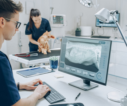
Their expertise in handling animals, positioning them correctly, and ensuring optimal image quality is essential for accurate diagnoses. Continuous Education Staying Updated with Advancements Veterinary radiography is a constantly evolving field. Poor positioning can lead to retakes, increased radiation exposure, and misdiagnoses.
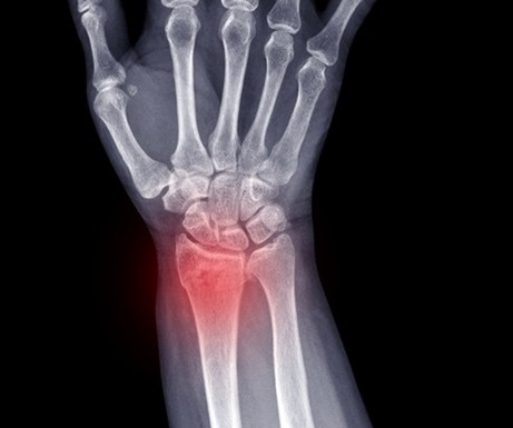
A new meta-analysis reveals that AI algorithms, especially CNNs, are highly effective in detecting wrist fractures from plain X-rays, performing on par with trained healthcare professionals.
A new computer-based diagnostic tool combines ultrasound imagery with specific clinical indicators to evaluate the risk of moderate-to-severe renal fibrosis.
Scientists have developed a sophisticated machine learning model that can more accurately predict the concussion status in patients and could be integrated into clinical practice.
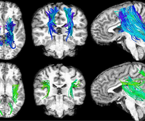
Morphological markers on MRI help diagnose autism. Shah, et al, Radiography , January 9, 2024. Image from Francisco Zamorano Mendieta, PhD, of the Universidad San Sebastián in Santiago, Chile Hypometabolism detected with F-18 FDG-PET in long-COVID patients : putative astrocyte dysfunction and glutamatergic dysregulation.
Introduction: The world of radiography is one filled with immense responsibility and precision, where the journey to capture images of the human body’s inner workings is not without its challenges. Radiation Safety: A Top Priority: Radiation safety is paramount in radiography.
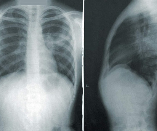
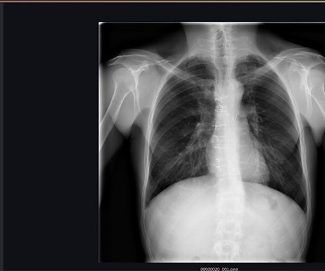
Teleradiology Introduction: The landscape of chest radiography is undergoing a revolutionary transformation, with deep learning at its forefront. This blog post explores how deep learning algorithms are reshaping the future of chest radiography, particularly in the realm of pneumonia detection and classification.
This annual event recognizes the hard work and dedication of radiologic technologists (RTs) who operate imaging technology to aid in accurate diagnoses and treatments. For instance, radiography reported an 18.1% With the demand for qualified RTs on the rise, hiring the right talent has never been more crucial for healthcare providers.
to demonstrate portable digital radiography and its effectiveness at high altitudes. Cairnie says, “Radiograph technology is critical for diagnosing several conditions, including bone fracture and chest conditions such as Tuberculosis. We were glad the expedition went well and now this technology is proven at high elevations."
. | W3-SSMK08 | Room 5E450A In this session on musculoskeletal imaging, a deep-learning AI algorithm will be presented for measuring shoulder kinematics using dynamic digital radiography (DDR) images. Check out this session on Tuesday morning for the details.
Over the years, advancements in diagnostic imaging have greatly increased patients’ overall care, quality of life, and outcome when diagnosed with certain conditions. Diagnostic imaging is an important tool used every day in healthcare to assist doctors in making the most informed decisions for their patients.
He said: "This innovative technology has the potential to change the way we diagnose a range of medical conditions. Advisory Board member, is also very impressed with the ARC60 imaging platform capabilities.
A research team exploring the potential use of standard ultrasound machines as an alternative to X-ray machines for diagnosing distal forearm fractures in children has found that ultrasound equipment can be a reliable diagnostic tool.
The demands on radiography are growing in number and complexity, and Carestream is dedicated to delivering solutions that help solve our customers’ challenges,” said Vincent Chan , president and general manager of digital radiography at Carestream.
Digital radiography utilizes a unique technology, similar to a digital camera, that instantly sends X-ray images to a computer for quick viewing. Likewise, digital radiography systems use a detector known as a flat panel to translate data into a digital electronic signal. This digital electronic signal becomes a digital X-ray image.
An AI algorithm was presented that could make dynamic digital radiography (DDR) more efficient by automatically measuring kinematics involved in certain shoulder injuries. Research from a group in Germany was presented suggesting that an AI algorithm for pneumothorax detection can perform similarly to radiology resident readers.
Key Points: Currently plain radiographs are the standard method in diagnosing syndesmotic ankle injuries even though the distal tibiofibular joint cannot be assessed due to superposition of the osseous structures in the foot.
A new study evaluated the effectiveness of gantry-free CBCT compared to two-dimensional radiography in diagnosing acute elbow trauma in both adults and children.
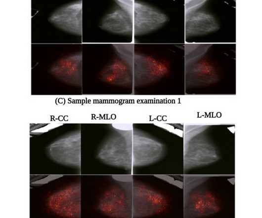
A new artificial intelligence-based algorithm uses deep learning to analyze multiple mammogram views concurrently, simulating the evaluation process of radiologists.
Chest radiography is a common diagnostic tool, but significant training and experience is required to interpret exams correctly,” said lead researcher Louis L. Too many false-positive diagnoses would result in unnecessary imaging, radiation exposure and increased costs.” Plesner, M.D., resident radiologist and Ph.D.
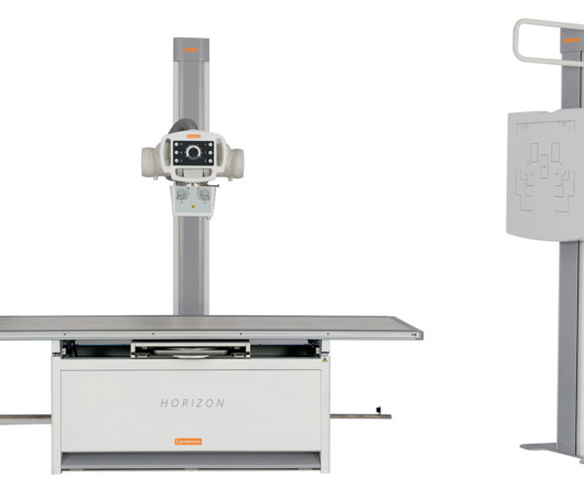
The intuitive and user-friendly manual analog system offers all the key functionality needed to help providers diagnose with confidence, provide exceptional patient care, and improve clinical outcomes. Delivering modern functionality in a compact, floor-mount design, the Horizon X-ray System is Carestream’s most affordable X-ray imaging room.
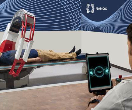
Nanox.ARC is a stationary X-ray system intended to produce tomographic images of the human musculoskeletal system adjunctive to conventional radiography on adult patients. Medical imaging is essential for detecting, diagnosing, and managing disease, guiding treatment decisions for improved health outcomes.
Yet a lack of access to high-quality imaging can lead to delayed or missed diagnoses, further exacerbating the health disparities experienced by people who live in disadvantaged or remote communities. Low-dose X-ray Solutions Serve Broadest Patient Population Digital radiography has come a long way at Massac Memorial as well.
Radiography soon became a vital tool in medicine, and for decades, it relied on photographic film to capture and develop X-ray images. The Birth of Digital Radiography: The 21st century brought a seismic shift as X-ray imaging transitioned from film to digital radiography.
From their serendipitous discovery to their pivotal place in diagnosing hidden conditions, we will illuminate the invisible to understand the lasting impact of X-rays. Chapter 3: The Evolution of Radiography: From Shadows to Images An exploration of the development of radiography, the earliest X-ray imaging technique.
From its early discovery to its integral position in diagnosing hidden conditions, we will uncover the layers beneath the surface to appreciate the indispensable role of X-ray imaging in patient care. How radiography transformed healthcare by allowing the visualization of internal structures.
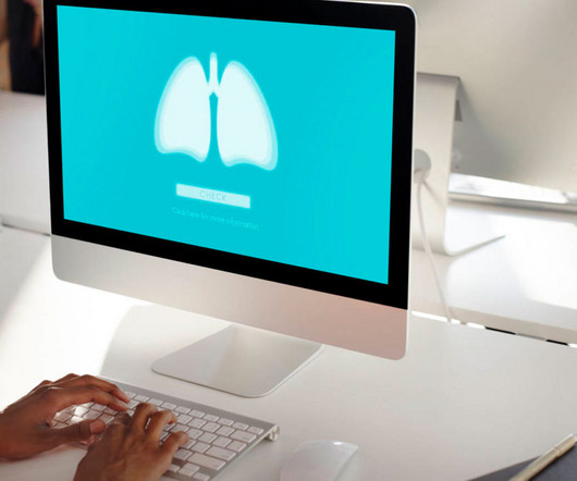
An innovative tool leverages the power of cognitive computing and deep learning to help radiologists and healthcare providers diagnose various chest conditions from X-ray images, thereby enhancing patient outcomes.
We organize all of the trending information in your field so you don't have to. Join 5,000 users and stay up to date on the latest articles your peers are reading.
You know about us, now we want to get to know you!
Let's personalize your content
Let's get even more personalized
We recognize your account from another site in our network, please click 'Send Email' below to continue with verifying your account and setting a password.


Let's personalize your content