This site uses cookies to improve your experience. To help us insure we adhere to various privacy regulations, please select your country/region of residence. If you do not select a country, we will assume you are from the United States. Select your Cookie Settings or view our Privacy Policy and Terms of Use.
Cookie Settings
Cookies and similar technologies are used on this website for proper function of the website, for tracking performance analytics and for marketing purposes. We and some of our third-party providers may use cookie data for various purposes. Please review the cookie settings below and choose your preference.
Used for the proper function of the website
Used for monitoring website traffic and interactions
Cookie Settings
Cookies and similar technologies are used on this website for proper function of the website, for tracking performance analytics and for marketing purposes. We and some of our third-party providers may use cookie data for various purposes. Please review the cookie settings below and choose your preference.
Strictly Necessary: Used for the proper function of the website
Performance/Analytics: Used for monitoring website traffic and interactions
Lung ultrasound (LUS) scoring is reliable for determining whether preterm newborns need surfactant for suspected respiratory distress syndrome (RDS), a study published February 19 in Pediatrics & Neonatology found. It also compared ultrasound scores with chest x-ray scores to predict the need for surfactant administration.
Lung ultrasound underdiagnoses clinically significant pneumothorax, suggest findings published September 20 in Surgery. Researchers led by Jarrett Santorelli, MD, from the University of California, San Diego found that ultrasound on initial trauma evaluation has low sensitivity and a high rate of false-negative exams.
While services for breast and lung cancer screening were temporarily halted, imagers in x-ray, lung ultrasound, and PET/CT were busy examining patients who presented with COVID-19. Medical imaging played a significant role in the early days of the pandemic when it hit its initial peak in April 2020.
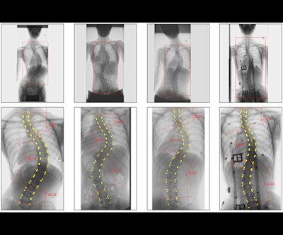
Orthopedic surgeons in Japan have developed an AI method based on spine x-ray. Read more on AuntMinnie.com Related Reading: Can an AI photo app help diagnose scoliosis? Is breast cancer surgery linked to spinal deformity?
It is often used to diagnose brain and spinal cord disorders, joint and musculoskeletal conditions, and cancer. X-RaysX-Rays are one of the most commonly and widely used diagnostic imaging techniques. X-RaysX-Rays are one of the most commonly and widely used diagnostic imaging techniques.
Diagnostic imaging tests are tools used by physicians to diagnose a range of medical conditions. For X-rays, it usually takes less than 10 minutes. An ultrasound can take about 30 minutes to an hour while CT scans can take 10-20 minutes. X-ray Also called a radiograph, an X-ray uses radiation to create images of the body.
12, 2025 Konica Minolta Healthcare Americas, has published a case study by clinicians in the pulmonary and radiology departments at ASST Fatebenefratelli Sacco (Milan, Italy) demonstrating the use of Dynamic Digital Radiography (DDR) to help definitively diagnose diaphragm dysfunction. tim.hodson Fri, 02/14/2025 - 15:14 Feb.12,
When Imaging Helps: Chest X-rays are the first-line imaging choice for diagnosing pneumonia, showing lung infections or fluid buildup. When Imaging Helps: X-rays can reveal joint space narrowing or bone changes due to arthritis. When Imaging Helps: X-rays are typically used to detect fractures or dislocations.
This was a case of the radiology practice miscoding an ultrasound exam conducted on the pregnant patient at 41 weeks that showed low amniotic fluid -- which resulted in the scan being sent "to nowhere," rather than to the mother's midwife. The baby died.
Morphological markers on MRI help diagnose autism. Ultrasound model predicts liver disease progression. Appropriateness and imaging outcomes of ultrasound, CT, and MR in the emergency department: a retrospective analysis from an urban academic center. Image from Eric Guedj, MD, PhD, of Marseille University Hospital, et al.
Future healthcare success will rely heavily on giving priority to key technologies like point-of-care ultrasound, 3D printing of anatomical organs, and artificial intelligence, RSNA 2023 attendees are set to find out during Tuesday's keenly anticipated international session on Singapore. Courtesy of Dr. Cher Heng Tan.
A research team exploring the potential use of standard ultrasound machines as an alternative to X-ray machines for diagnosing distal forearm fractures in children has found that ultrasound equipment can be a reliable diagnostic tool.
A team of doctors, nurses and medical researchers affiliated with multiple institutions in Australia has found that ultrasound devices can serve as reliable diagnostic tools for children presenting symptoms of distal forearm fracture.
A research team exploring the potential use of standard ultrasound machines as an alternative to X-ray machines for diagnosing distal forearm fractures in children has found that ultrasound equipment can be a reliable diagnostic tool.
milla1cf Thu, 03/21/2024 - 10:42 March 21, 2024 — Artificial intelligence can spot COVID-19 in lung ultrasound images much like facial recognition software can spot a face in a crowd, new research shows. How does AI analyze ultrasound lung images?
Portable ultrasound devices could provide an alternative to x-ray machines for diagnosing forearm fractures in children in a move that could alleviate waiting times for families in hospital emergency departments (ED).
Radiologists are medical doctors who specialize in interpreting imaging studies like X-rays, CT scans, MRIs, and ultrasounds to diagnose and guide treatment for various conditions. This rigorous training covers all imaging modalities, from X-rays to advanced techniques like MRI and PET/CT scans.
“X-Rays and Beyond” embarks on an exciting journey to explore the captivating world of radiology, highlighting how it goes far beyond just X-rays. The Discovery of X-Rays: Introduce the pioneering discovery of X-rays by Wilhelm Conrad Roentgen in 1895 and how it revolutionized medicine.
The list below are imaging exams used to diagnose breast cancer. During a diagnostic mammogram, multiple x-rays are taken at different angles so that views of a specific area of interest can be further examined. Ultrasound Breast ultrasounds use sound waves to look inside your breasts.
It assists with diagnosing the presence of tumors and determining if the cancer has spread. To help guide the diagnostic process, the following imaging procedures may be used: X-Ray: Typically the first requested imaging procedure, X-rays help identify respiratory abnormalities and other lung issues.
Over the years, advancements in diagnostic imaging have greatly increased patients’ overall care, quality of life, and outcome when diagnosed with certain conditions. Diagnostic imaging is an important tool used every day in healthcare to assist doctors in making the most informed decisions for their patients.
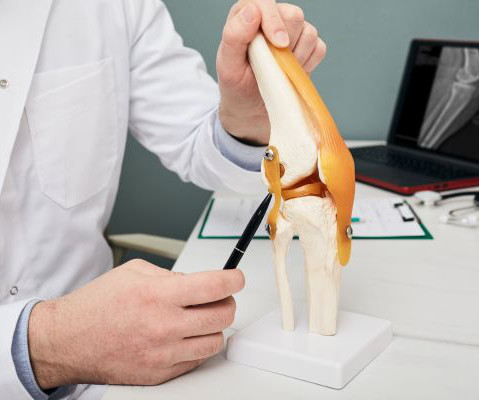
Diagnosing an ACL Injury with an MRI For ACL injuries, the diagnostic process starts with a physical exam. MRI is the primary tool, although an X-ray may be ordered to rule out broken bones and an ultrasound to check for the extent of internal injuries. Contact Midstate Radiology Associates to make an appointment today.
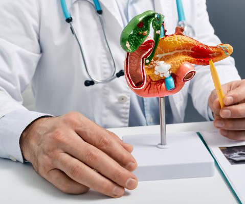
This procedure is less invasive than other methods and provides a higher level of detail compared to traditional X-rays, while also reducing radiation exposure. Ultrasound: An abdominal or endoscopic ultrasound can reduce radiation exposure, while obtaining images of the pancreas.
Medical imaging via X-rays, CT scans, MRIs and ultrasounds provide health-care professionals with unique perspectives and a better understanding of what's happening inside a patient's body. Using various forms of waves, these machines can visualize many unseen ailments and diseases.
Medical imaging plays a pivotal role in diagnosing and monitoring various medical conditions, but navigating the associated costs can be challenging. Medical Imaging and Its Importance: Medical imaging encompasses a wide range of technologies like X-rays, CT scans, MRI, and ultrasound.
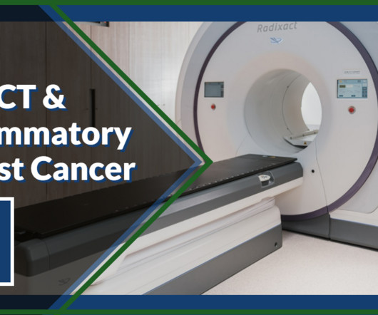
But cancer in initially diagnosed IBC patients has already metastasized about 20 percent of the time, approximately doubling the percentage of other breast cancer patients. Why is PET/CT Critical in Diagnosing IBC? IBC patients aren’t always diagnosed with a palpable mass, so it’s difficult to target a biopsy.
One of the most exciting of these new frontiers is ultrasound technology. With its ability to safely and painlessly penetrate the human body, scientists and doctors are discovering new possibilities in diagnosing and treating conditions that previously required more invasive procedures.
It encompasses various imaging techniques, including X-rays, CT scans, MRIs, and ultrasounds, allowing medical professionals to peer inside the body and diagnose a wide range of conditions. Radiology plays an unseen but vital role in modern healthcare.
In this blog post, we’ll explore the differences and uses of MRI, CT scans, X-rays, ultrasounds, and PET/CT to help you better understand what to expect and how these technologies can assist in your healthcare. Unlike X-rays and CT scans, MRIs do not use ionizing radiation.
State-of-the-Art Technology Not all imaging centers offer the same equipment, and newer technology often leads to more precise imaging and quicker diagnoses. Ensuring your imaging center has these certifications guarantees that your scans will be handled by knowledgeable and experienced professionals.
You now have access to high-quality, low-cost ultrasound in King George at our newest outpatient facility. Located at 11131 Journal Pkwy, Medical Imaging of King George offers ultrasound imaging services by appointment Monday through Friday from 8:30 a.m. To schedule your ultrasound in King George, call 540-741-9729 (XRAY).
Mobile imaging provides comprehensive X-Ray, EKG and ultrasound services directly to medical facilities,homesand businesses. When people hear the world mobile imaging, they typically think of portable X-Ray imaging. What is Mobile Imaging? Unfortunately, each of thesescenarios comes with a drawback.
Medical imaging is a powerful tool for diagnosing and treating medical conditions; however, it can also expose patients to excess radiation levels. Technicians have designed these machines with unique components that limit X-ray emissions while still providing high-quality imaging.
Careverse's products cover various modalities of medical imaging, such as CT, MRI, ultrasound, X-rays and mammography, etc., This significantly reduces the manual workload of radiologists, enabling them to dedicate more time to accurate diagnoses and seamless collaboration with other clinical departments.
In one of our studies, we took tissue from patients with NSF and did electron microscopy with x-ray dispersion analysis to try to determine whether gadolinium was actually in the tissue that was biopsied. Sadowski said that MR can be avoided altogether by using ultrasound, CT, or molecular imaging.
Radiology plays a pivotal role in diagnosing and managing these injuries, providing valuable insights that guide treatment strategies and facilitate the prompt return of athletes to their respective fields. Discuss how X-rays are crucial for identifying fractures, dislocations, and other acute skeletal injuries on the field.
At PURE Mammography, we offer 3D breast mammography as well as ultrasound imaging for breasts and other areas of the body. In addition to mammograms and ultrasounds, women should know about the various other breast screenings that are performed. Breast Ultrasound A breast ultrasound is different than an x-ray.
Doctors use imaging tests to see inside a patient’s body and diagnose their illnesses and injuries. There are several types of imaging tests that physicians use to detect cancer in patients: X-Ray, Computed Tomography (CT), Magnetic Resonance Imaging (MRI), Ultrasound (US), Nuclear Medicine, and Positron Emission Tomography (PET).
Radiology is a medical imaging procedure that uses ionizing electromagnetic radiation to create images of bones, organs, and soft tissues to diagnose a patient’s symptoms, disease, or conditions. It includes techniques like X-rays, CT scans, MRIs, ultrasounds, and fluoroscopy.
Whether it’s through X-ray, MRI, CT scan, ultrasound, or using tiny cameras, health care professionals can see beyond the flesh and gain a deeper understanding of the human body’s internal machinations. These imaging methods diagnose all kinds of conditions, but can often be intimidating.
Mobile imaging provides comprehensive X-Ray, EKG and ultrasound services directly to medical facilities, homes and businesses. When people hear the world mobile imaging, they typically think of portable X-Ray imaging. What is Mobile Imaging? Unfortunately, each of these scenarios comes with a drawback.
Although the word “radiology” sounds like it involves radiation, that is not always the case – for example, MRI (magnetic resonance imaging) and ultrasound do not use radiation in their medical imaging technologies. Digital X-rays use less radiation and are employed for the same purposes.
Medical imaging is used to find, diagnose, monitor, and even treat different medical conditions or injury. Take x-rays for example. X-rays are used to view the skeletal system – the bones – of the body. X-rays are used to identify different issues with a patient’s bones and joints.
We organize all of the trending information in your field so you don't have to. Join 5,000 users and stay up to date on the latest articles your peers are reading.
You know about us, now we want to get to know you!
Let's personalize your content
Let's get even more personalized
We recognize your account from another site in our network, please click 'Send Email' below to continue with verifying your account and setting a password.


Let's personalize your content