This site uses cookies to improve your experience. To help us insure we adhere to various privacy regulations, please select your country/region of residence. If you do not select a country, we will assume you are from the United States. Select your Cookie Settings or view our Privacy Policy and Terms of Use.
Cookie Settings
Cookies and similar technologies are used on this website for proper function of the website, for tracking performance analytics and for marketing purposes. We and some of our third-party providers may use cookie data for various purposes. Please review the cookie settings below and choose your preference.
Used for the proper function of the website
Used for monitoring website traffic and interactions
Cookie Settings
Cookies and similar technologies are used on this website for proper function of the website, for tracking performance analytics and for marketing purposes. We and some of our third-party providers may use cookie data for various purposes. Please review the cookie settings below and choose your preference.
Strictly Necessary: Used for the proper function of the website
Performance/Analytics: Used for monitoring website traffic and interactions
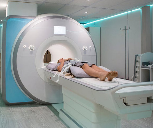
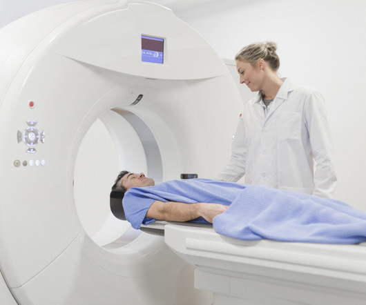
The Power of DiagnosticImaging in Early Disease Detection Medical imaging is one of the most significant developments in medical science. By allowing doctors to view the inside of the body without the need for surgery, this technology aids in the early detection of diseases. What is DiagnosticImaging?
DiagnosticImaging is a great tool for your medical professional to use to detect issues sooner rather than later. There are several types of diagnosticimaging available today; each one used to visualize the internal structures of the body to assist doctors in diagnosis and treating various diseases and medical conditions.
and radiology has learned much since then, according to experts who directly dealt with the diseases impact. While the pandemic affected medical operations across the country, the experts said that radiologists developed and honed their sense of resiliency as imaging was placed on the front lines. But that involves human contact.
Eliot Siegel, MD; Stanislav Spiridonov, MD; Nathan Gee, MD; and Anthony Chang, PhD, are among a niche gathering of early adopters, entrepreneurial physicians, medical physicists, and investors with a sweet spot for nuclear medicine, diagnostic radiology, and radiation oncology.
However, increased longevity has brought on a troubling rise in chronic disease, diminishing the quality of life later in life. Centers for Disease Control and Prevention (CDC) found that more than a quarter of U.S. Mark Crockett, MD, chief medical officer at TeleDaaS. Americans today can expect to live long lives.
His research interests include using structural and functional MRI -- particularly ultrahigh-field, 7-tesla MRI -- to map brain microstructure and develop neurosurgical treatment of brain tumors, epilepsy, and neurodegenerative and movement disorders such as Parkinson's disease, essential tremor, and dystonia. Elliot Fishman, MD.
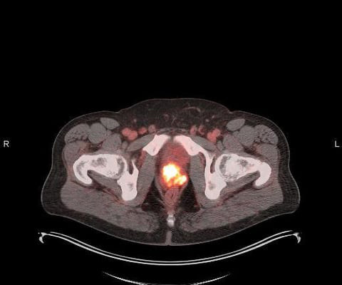
Theranostics pairs diagnostic biomarkers that can be visualized on nuclear medicine imaging with therapeutic agents that share a specific target in diseased cells or tissues. After binding to the receptor, the drug works by entering the cell allowing radiation to cause damage to the tumor cells.
Deep learning-based image reconstruction (DLR) has been a hot topic in CT over the past five years, as researchers and vendors have continuously demonstrated the technology's potential to improve on legacy and filtered back-projection (FBP) reconstruction methods. Accordingly, commercial and research activity has accelerated.
While the medical imaging technique is widely used and is critical for the early detection of cancers and diseases, the survey revealed that many Canadians hold harmful misconceptions about MRIs that may deter them from getting screened. While very high radiation doses can damage or kill eggs or sperm, MRI scans are radiation-free.
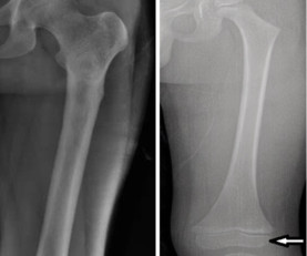
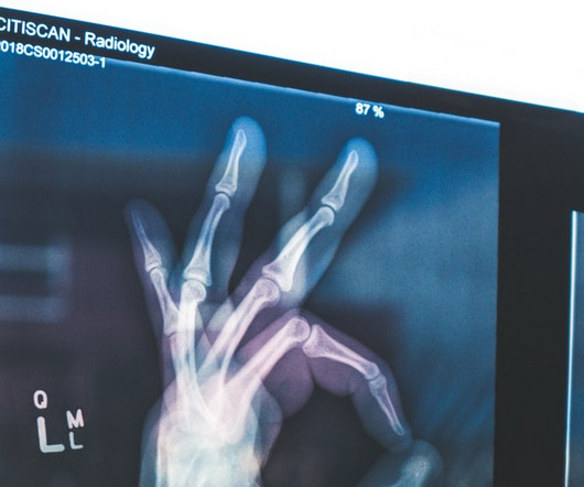
Here’s how: DiagnosticImaging : X-rays provide detailed images of internal structures such as bones, organs, and tissues. They help diagnose fractures, identify abnormalities in organs, detect tumors, and assess the extent of injuries or diseases.
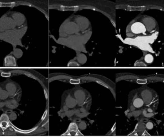
CT scans also aid in surgical planning as well as monitoring the effectiveness of cancer treatments like chemo or radiation therapy. Evaluating the Tissue and Organs in the Chest Chest CT scans are are more detailed than x-rays, giving you more information about possible diseases or injury of your chest organs.
Emerging research on coronary artery calcium scoring for the assessment of coronary artery disease (CAD) suggests the use of virtual non-contrast images from photon-counting CT may lead to a nearly 20 percent reduction in radiation dosing.
milla1cf Mon, 05/15/2023 - 10:26 May 15, 2023 — GE HealthCare is presenting three new global innovations – Intelligent Radiation Therapy (iRT), Auto Segmentation, and an updated Magnetic Resonance (MR) Radiation Therapy Suite (AIR Open Coil Suite) – that underscore the company’s commitment to enhancing the radiation oncology care pathway.
The Early Signs of Breast Cancer Breast cancer is a disease where abnormal cells divide without control in one or both of your breasts. The earlier breast cancer is detected through diagnosticimaging, the better chance there is for successful treatment with surgery, radiation therapy, or chemotherapy. Now is the time.
GE HealthCare expects to leverage MIM Software’s imaging analytics and digital workflow capabilities across various care areas to accelerate innovation and differentiate its solutions for the benefit of patients and healthcare systems around the world.
Point of Care Ultrasound (POCUS) is an ultrasound-based diagnostic tool used in clinical settings that uses sound waves to create images allowing doctors to see inside the body without having to do surgery or other invasive procedures. For more information about Vesta’s teleradiology services, please contact us today.
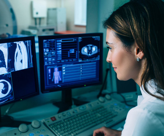
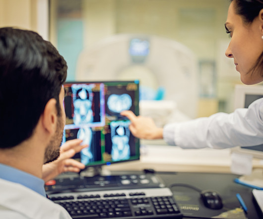
milla1cf Thu, 08/24/2023 - 16:02 August 24, 2023 — Medical imaging via X-rays , CT scans , MRIs and ultrasounds provide health-care professionals with unique perspectives and a better understanding of what’s happening inside a patient’s body. Using various forms of waves, these machines can visualize many unseen ailments and diseases.
Theranostics pairs diagnostic biomarkers that can be visualized on nuclear medicine imaging with therapeutic agents that share a specific target in diseased cells or tissues. After binding to the receptor, the drug works by entering the cell allowing radiation to cause damage to the tumor cells.
Intelligent imaging systems that learn with each scan and produce medical images with unprecedented speed, detail, and precision represent some of the most seismic advances in radiology since the Nobel Prize in Medicine was awarded to the inventors of computer-assisted tomography in 1979. Kelly Londy of GE HealthCare.
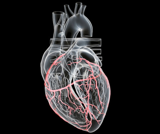
CT scans also aid in surgical planning as well as monitoring the effectiveness of cancer treatments like chemo or radiation therapy. By producing interior images of the coronary arteries, CT scans help identify blockages, aneurysms, or other abnormalities that can lead to damaging issues like a heart attack or stroke.
Photon-counting CT enables the acquisition of more detailed images with anatomical and functional information by “counting” each individual X-ray photon that passes through patients. Thus, without increasing the radiation dose, small abnormalities become visible.
Medical professionals also discovered the inherent radiation risks with the process and have now improved the protocols and techniques to minimize this exposure. With the use of some ionizing radiation, a clear picture is produced and a physician can find the precise location of a fracture. Pinpointing injuries. Mammography.
The radiohybrid PSMA theranostic technology platform enables molecules within the class to be modified and deployed for either diagnostic PET imaging or therapeutic applications, and can also be developed with both beta- and alpha-emitting therapeutic radioisotopes. higher tumor absorbed radiation dose. than 177Lu-PSMA-I&T.
i Thankfully, the field of breast screening is not static, and advancements are revolutionizing how we detect and diagnose the disease. J Med ImagingRadiat Sci. DiagnosticImaging Utilization in the Emergency Department: Recent Trends in Volume and Radiology Work Relative Value Units. doi: 10.1016/j.jmir.2023.10.001.
produced, non-uranium based Mo-99 for use in diagnosticimaging. These capabilities will help them develop and deliver products with the potential to improve care for even more patients with serious disease. “In In parallel with these initiatives, NorthStar’s imaging portfolio continues to advance,” Mr. Merrick further added.
The balance of dose and image quality is even more important in pediatric medical imaging. Not only are children more radiosensitive than adults (the cancer risk per unit dose of ionizing radiation is higher), but children also have a longer expected lifetime, which puts them at greater risk of cancer following radiation exposure.(1)
milla1cf Tue, 05/02/2023 - 23:50 May 2, 2023 — Blue Earth Diagnostics , a Bracco company and recognized leader in the development and commercialization of innovative PET radiopharmaceuticals, today announced additional results from its completed Phase 3 SPOTLIGHT trial of 18F-rhPSMA-7.3 18F-rhPSMA-7.3 on behalf of the SPOTLIGHT Study Group.
It represents a new class of purposely engineered, high-affinity PSMA-targeted radiopharmaceuticals based on novel radiohybrid technology, which may offer diagnosticimaging and therapeutic potential. POSLUMA was developed to assist physicians in the detection and localization of prostate cancer. POSLUMA was approved by the U.S.
In this blog post, we’ll explore the perils associated with inadequate interpretation of diagnosticimaging and the potential risks it poses to patients. Diseases such as cancer, when overlooked, can progress to advanced stages with poorer prognoses.
POSLUMA represents a new class of high-affinity PSMA-targeted PET radiopharmaceuticals based on novel radiohybrid technology and is labeled with the radioisotope 18F to provide readily available patient access and leverage the high image quality of 18F-labeled PSMA PET imaging to facilitate effective detection of disease.
Radiology is a branch of medicine that uses radiant energy in the diagnosis and treatment of disease. Practitioners of radiology are called radiologists, and they utilize imaging technology in the diagnosis and treatment of patients. Medical imaging is a technology which is used by radiologists , particularly for diagnostic purposes.
Chapter 2: Basics of X-ray Science – A Radiologic Primer A detailed explanation of the scientific principles that govern X-ray imaging. How X-rays are generated, interact with human tissue, and create diagnosticimages. The technical precision and operation of equipment to produce high-quality diagnosticimages.
Discuss how theranostic approaches integrate diagnosticimaging and targeted therapy for more personalized and effective treatments. Discuss how this approach delivers radiation with exceptional precision to cancer cells. Discuss how these compounds are expanding therapeutic options for various diseases, including cancer.
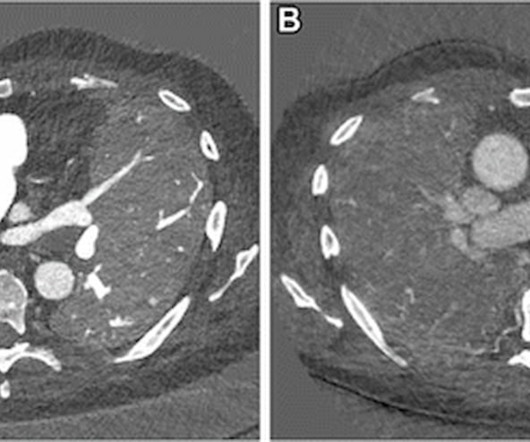
For conditions ranging from interstitial lung disease and post-COVID-19 complications to idiopathic pulmonary fibrosis, photon-counting computed tomography (PCCT) facilitates simultaneous functional and morphologic lung assessment at lower radiation dosing than conventional chest CT, according to newly published research.
Pediatric Radiology is a subspecialty of radiology that deals with the imaging of infants, children, and adolescents. Children and young adults under 21 have different anatomy, disease processes, and emotional and physical needs when compared to adult patients.
A radiology department is responsible for providing diagnostic studies and radiation therapy. Because they work with radiation, there are radiation exposure risks associated with any job in radiology. Some common radiology studies include x-rays, CT scans, MRIs, mammography, and ultrasound.
Gum Health Assessment: Periodontal disease often starts beneath the gumline. Dental X-rays, like periapical and panoramic X-rays, provide insights into bone loss and gum health, enabling the early diagnosis and management of gum diseases like periodontitis. This aids in diagnosing hidden sources of pain or discomfort.
The historical context of Wilhelm Roentgen’s discovery and the birth of this revolutionary diagnostic tool. Chapter 2: How X-rays Work: Illuminating the Invisible A detailed explanation of the science behind X-ray imaging. The principles of radiation and how X-rays interact with the human body to create diagnosticimages.
Thanks to new diagnostic approaches, patients can be grouped according to the biomarkers identified through imaging, providing a deeper understanding of the molecular basis of their disease and the appropriate course of treatment. In addition, the combination of PET/MRI imaging is proving to be even more powerful than MRI alone.
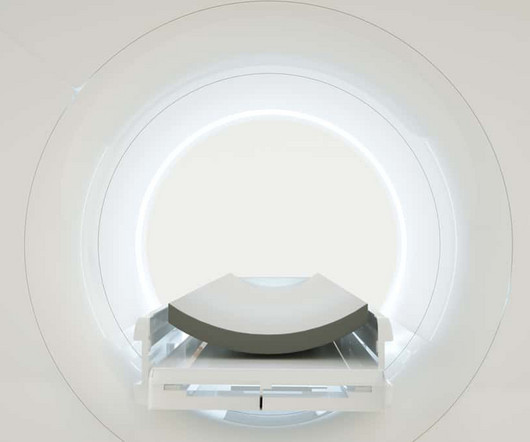
Diagnosticimaging plays a very important role in modern medicine. It allows physicians to diagnose and treat many different health conditions, injuries, and diseases more accurately and easily. A CT (computerized tomography) scan is used to enable doctors to see clear images of the structures and tissues of your body.
Explore how precision imaging agents are enhancing diagnostic accuracy and enabling the visualization of specific molecular targets. Discuss how theranostic approaches combine diagnosticimaging and targeted therapy, allowing for personalized and precise treatment strategies.
Effective initial staging of prostate cancer, particularly with regards to the detection of metastatic disease, is critical to optimal clinical management of patients,” said Phillip H. Kuo, MD, Ph.D. , Departments of Medical Imaging, Medicine, and Biomedical Engineering. Blue Earth Diagnostics received U.S.
Introduction: The journey from 2D to 3D imaging in dental and medical practices has been marked by the advent of Cone Beam Computed Tomography (CBCT). This transformative technology has taken diagnosticimaging to new heights, providing insights that transcend the limitations of traditional two-dimensional methods.
Osteoporosis is a common bone disease with as many as 1.5 The scan uses a very low dose of radiation to produce pictures of the inside of the body, particularly around the hips and lower spine, to measure bone mass and strength. This weakens bones making them porous, fragile, and susceptible to fractures.
We organize all of the trending information in your field so you don't have to. Join 5,000 users and stay up to date on the latest articles your peers are reading.
You know about us, now we want to get to know you!
Let's personalize your content
Let's get even more personalized
We recognize your account from another site in our network, please click 'Send Email' below to continue with verifying your account and setting a password.


Let's personalize your content