This site uses cookies to improve your experience. To help us insure we adhere to various privacy regulations, please select your country/region of residence. If you do not select a country, we will assume you are from the United States. Select your Cookie Settings or view our Privacy Policy and Terms of Use.
Cookie Settings
Cookies and similar technologies are used on this website for proper function of the website, for tracking performance analytics and for marketing purposes. We and some of our third-party providers may use cookie data for various purposes. Please review the cookie settings below and choose your preference.
Used for the proper function of the website
Used for monitoring website traffic and interactions
Cookie Settings
Cookies and similar technologies are used on this website for proper function of the website, for tracking performance analytics and for marketing purposes. We and some of our third-party providers may use cookie data for various purposes. Please review the cookie settings below and choose your preference.
Strictly Necessary: Used for the proper function of the website
Performance/Analytics: Used for monitoring website traffic and interactions
partnership that will begin by upgrading and replacing cancer-specific diagnosticimaging equipment. The investment involves replacing existing imaging equipment at the end of its life cycle with AI-enabled treatment equipment, according to the Alberta government.
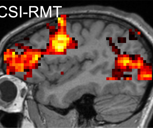
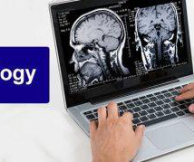
NYU Langone Health spinout Microstructure Imaging (MICSI) has secured U.S. Food and Drug Administration (FDA) 510(k) clearance for its MICSI-RMT AI imageenhancement software for brain MRI. Designed to enhance white-matter imaging, MICSI-RMT employs a random matrix theory-based algorithm.
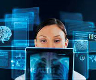
Among the most vital tools in this preventive health is diagnosticimaging. Professional Radiology in El Paso is a top diagnosticimaging center. We are known for our accurate and detailed imaging, which ensures the best possible care for our patients. Call (915) 225-2480 today to schedule an appointment.
AI also can help with specific elements of imageenhancement, such as automatically removing or suppressing the appearance of bone to enhance soft-tissue visualization. Many attendees were looking for ways AI can enable imaging teams to drive bottom-line efficiencies.
Key opinion leaders explore the significance of interdisciplinary collaboration in prostate cancer management, with a focus on how PSMA PET imagingenhances decision-making processes within multidisciplinary care teams.
Chapter 1: The Digital Jigsaw – Understanding CT Image Reconstruction: Begin by unraveling the concept of CT image reconstruction, how it works, and its significance in creating detailed diagnosticimages.
Introduction: Radiologists are the artists in the realm of diagnosticimaging, using CT (Computed Tomography) scans as their canvas to craft diagnostic masterpieces. Explore their collaborative efforts with physicians, CT technologists, and other healthcare professionals to ensure accurate diagnoses.
Science in Action: The X-ray Imaging Process: At its core, X-ray imaging is a precise scientific process. It involves the emission of X-ray photons, their interaction with tissues of varying densities, and the capture of transmitted X-rays to create diagnosticimages.
Introduction: In the world of diagnosticimaging, CT (Computed Tomography) scans are the rays of insight that have transformed patient care. This blog explores the pivotal role that CT scans play in healthcare, providing a closer look at how they have reshaped the diagnostic landscape.
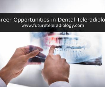
High-Resolution Imaging Modalities: Highlight the advent of high-resolution imaging modalities in teleradiology. Discuss how technologies like high-field MRI, multi-slice CT, and digital mammography contribute to clearer and more detailed diagnosticimages.
Advanced Imaging Modalities: Pushing Boundaries of Precision: Highlight the emergence of advanced imaging modalities such as high-resolution MRI and advanced CT scans. Discuss how these technologies provide unparalleled precision in diagnosticimaging.
The Canvas of Digital Images: Discuss the shift from traditional radiological films to the canvas of digital images in teleradiology. Explore how advancements in pixel resolution and digital rendering contribute to the clarity and precision of diagnosticimages.
As technology advances and healthcare systems embrace digital transformation, teleradiology is poised to play a pivotal role in shaping the future of diagnosticimaging. Enhanced visualization tools will provide more detailed and dynamic insights into medical images, aiding in comprehensive diagnostics.
Chapter 3: The Diagnostic Blueprint – How CT Scans are Constructed: Learn about the construction process of CT scans, exploring how raw data is transformed into diagnosticimages using mathematical algorithms and computational power.
Enhanced Accuracy and Precision: Explore how teleradiology contributes to enhanced accuracy and precision in diagnosticimaging. Integration of Advanced Imaging Modalities: Highlight how teleradiology integrates advanced imaging modalities.
The program plans to utilize AI algorithms to analyze radiology images, enhancing the accuracy and efficiency of cancer screening. The integration of AI in teleradiology will help automate image interpretation, expedite diagnoses, and reduce the workload on radiologists.
Swift transmission of diagnosticimagesenhances emergency preparedness. Teleradiology solutions are seamlessly integrated into the cultural fabric, fostering a harmonious blend of tradition and technology. **7.
We organize all of the trending information in your field so you don't have to. Join 5,000 users and stay up to date on the latest articles your peers are reading.
You know about us, now we want to get to know you!
Let's personalize your content
Let's get even more personalized
We recognize your account from another site in our network, please click 'Send Email' below to continue with verifying your account and setting a password.


Let's personalize your content