This site uses cookies to improve your experience. To help us insure we adhere to various privacy regulations, please select your country/region of residence. If you do not select a country, we will assume you are from the United States. Select your Cookie Settings or view our Privacy Policy and Terms of Use.
Cookie Settings
Cookies and similar technologies are used on this website for proper function of the website, for tracking performance analytics and for marketing purposes. We and some of our third-party providers may use cookie data for various purposes. Please review the cookie settings below and choose your preference.
Used for the proper function of the website
Used for monitoring website traffic and interactions
Cookie Settings
Cookies and similar technologies are used on this website for proper function of the website, for tracking performance analytics and for marketing purposes. We and some of our third-party providers may use cookie data for various purposes. Please review the cookie settings below and choose your preference.
Strictly Necessary: Used for the proper function of the website
Performance/Analytics: Used for monitoring website traffic and interactions
A dedicated teacher and radiologic technologist since 2009, Stewart currently serves as associate professor of diagnosticimaging, and program director of radiologic sciences at Quinnipiac University in Connecticut. I'm a radiographer,' " Stewart recalled. For example, in patientcare courses there is a natural crossover.
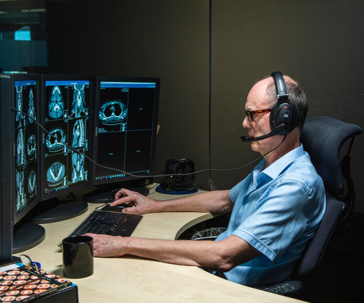
In any case, for Pickhardt, research is key to providing high-quality patientcare. "I I love how research improves my clinical acumen and 'moves the needle' for patientcare," he said. His area of expertise is neuroradiology, with a special emphasis on diagnosticimaging of the head, neck, brain, and spine.
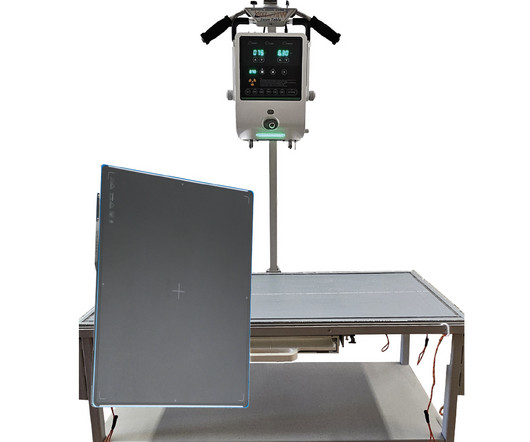
The system is a versatile, digital radiographic X-ray suite powered by Fujifilm’s latest FDR D-EVO III high-resolution, high sensitivity ultra-lightweight, glass-free detectors coupled with Dynamic Visualization , the company’s intelligent image processing. “At
The Impact on Patients and Clinical Management A 2024 study in the Journal of Clinical Imaging found that 68% of surveyed radiology practices had unreported radiology exams. At seven days post-imaging, nearly half had unreported brain and chest CT scans, while 59% had unreported chest radiographs.
Teleradiology-India Introduction: “X-ray Visionaries” takes you on a compelling journey to unveil the expertise of radiographers and technologists, the unsung heroes of X-ray technology. Chapter 1: Introduction to Radiographers and Technologists An overview of the pivotal roles radiographers and technologists play in healthcare.
But at RSNA 2023, part of the focus was on AI advances that are changing how radiographers work today—for example, continuing to help us balance the goal of capturing the most information possible in an image without excessive radiation dose. AI tools that enable radiographers to separate noise from an image already exist today.
ASRT President Brandon Smith, MBA If passed, MARCA would ensure complete Medicare reimbursement for services provided by a radiologist assistant, when supervised by a radiologist as part of a radiologist-led patientcare team, regardless of the setting in which the service is performed, according to the ASRT.
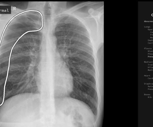
But how will AI in the workplace affect the radiographer and how does it differ from the red dot system radiographers are so familiar with? The Red Dot System Often one of the first courses a newly qualified radiographer attends is the red dot course. Image courtesy of qure.ai Here, the explanation is straightforward.
Kim Mason Kim Mason, an Audit and Research Radiographer for Mid Yorkshire Teaching Hospitals Trust, talks about their role as well as the value of radiographer engagement in research activities and how to get involved. Hi, I’m Kim and I am an alternative-styled, funky-haired, septum-pierced, disabled Audit and Research Radiographer.
The purpose of this blog is not to define requirements for reporting safety incidents, but rather to encourage and challenge radiology imaging directors to examine the culture and awareness around reporting within your departments. How are radiographers treated when they admit to making a mistake? Do they know who to contact?
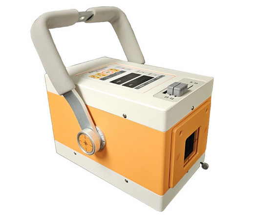
In the ever-evolving landscape of veterinary medicine, technological advancements continue to play a pivotal role in enhancing patientcare and diagnostic capabilities. One such innovation that has transformed the way veterinarians approach imaging is the evolution of portable x-ray units.
They might not be aware of time-saving features like Modality Performed Procedure Step, Custom Configurations or Automated Workflow; or all the tools available to help capture diagnosticimages at the highest quality possible. Start the new year off by assessing who in your department could benefit from more training.
At DiagnosticImaging Systems, we offer the Mixed Animal X-Ray Package , featuring state-of-the-art components such as the Elite 125100 Portable X-Ray Machine , the DR Wizard X 14×17 Wireless Digital Radiography Detector , and the VV200 Mobile X-Ray Table.
With advance planning and an emphasis on learning, your facility can perform imaging studies with the same standard of care as a large children’s hospital. Typically, one of two things may happen when an imaging technologist or sonographer who infrequently performs pediatric diagnosticimaging studies images a child.
Any time of the day or night, a clinician, radiographer, or radiology manager can call TMC to discuss scanning a patient. These can be emergency patients in the day or night, they can be acute inpatients who simply need that next step in their pathway or to be discharged safely, or perhaps just a routine scan which feels urgent.
DiagnosticImaging: Obstetrics, Elsevier, 2016, pp. Jay Vora is a medical student at Edward Via College of Osteopathic Medicine (VCOM) – Virginia and plans to pursue a residency in diagnostic radiology. Seeing the radiographicimages made medical education come to life for him. Russell, Sarah A.,
Will the technological leaps of the coming years eclipse the uniquely human capacities of empathy, judgment, and creativity that physicians bring to patientcare, or can we harness them to enhance -- and not eliminate -- our profession? DiagnosticImaging. RadioGraphics. J Am Coll Radiol. 2021;18(4):511-519.
Use of low-field MRI for this indication would improve patientcare by expanding access to the modality, wrote a team led by medical student Lauren Kelsey. The group's commentary was published February 13 in RadioGraphics. tesla or 3-tesla), identifying entities that may be adequately imaged at 0.55-tesla
The integration of AI is set to enhance radiologists' roles, granting them more control over their workday to focus on delivering excellent patientcare. The shortage of radiographers: A global crisis in healthcare. J Med Imaging Radiat Sci. 2023 Oct 19:S1939-8654(23)01877-5. doi: 10.1016/j.jmir.2023.10.001. 2023.10.001.
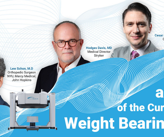
In the world of orthopedic diagnostics, imaging plays a crucial role in identifying deformities and planning surgical interventions. Conventional radiographs and MRIs have been the standard, but they come with limitations when it comes to understanding the complex, three-dimensional structure of the human body. Why Choose WBCT?
We organize all of the trending information in your field so you don't have to. Join 5,000 users and stay up to date on the latest articles your peers are reading.
You know about us, now we want to get to know you!
Let's personalize your content
Let's get even more personalized
We recognize your account from another site in our network, please click 'Send Email' below to continue with verifying your account and setting a password.


Let's personalize your content