This site uses cookies to improve your experience. To help us insure we adhere to various privacy regulations, please select your country/region of residence. If you do not select a country, we will assume you are from the United States. Select your Cookie Settings or view our Privacy Policy and Terms of Use.
Cookie Settings
Cookies and similar technologies are used on this website for proper function of the website, for tracking performance analytics and for marketing purposes. We and some of our third-party providers may use cookie data for various purposes. Please review the cookie settings below and choose your preference.
Used for the proper function of the website
Used for monitoring website traffic and interactions
Cookie Settings
Cookies and similar technologies are used on this website for proper function of the website, for tracking performance analytics and for marketing purposes. We and some of our third-party providers may use cookie data for various purposes. Please review the cookie settings below and choose your preference.
Strictly Necessary: Used for the proper function of the website
Performance/Analytics: Used for monitoring website traffic and interactions
The Power of DiagnosticImaging in Early Disease Detection Medical imaging is one of the most significant developments in medical science. What is DiagnosticImaging? Diagnosticimaging includes different types of scans that take pictures of the inside of the body.
DiagnosticImaging is a great tool for your medical professional to use to detect issues sooner rather than later. There are several types of diagnosticimaging available today; each one used to visualize the internal structures of the body to assist doctors in diagnosis and treating various diseases and medical conditions.
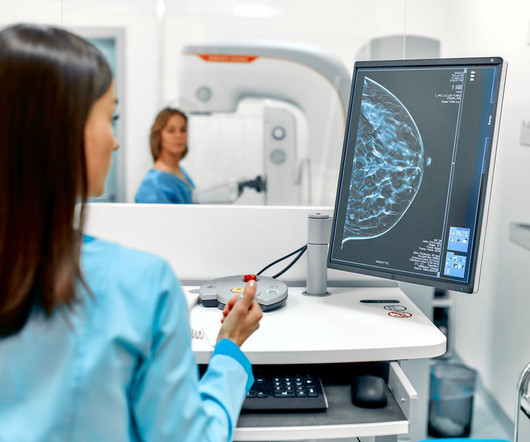
However, many patients have concerns about radiation exposure and the potential risks involved. The truth is mammograms are generally safe when used properly, and the amount of radiation you’re exposed to is minimal. As the leading diagnosticimaging radiology center in El Paso, patient care and safety are our top priorities.
While the use of ultrasound for pediatric appendicitis imaging has increased, the modality -- as well as MRI -- remains underutilized in this area, according to research published August 22 in the Journal of Pediatric Surgery. Ultrasound alone was used in 53.3% Ultrasound alone was used in 53.3% NSQIP-P hospital 71.7%
Medical imaging played a significant role in the early days of the pandemic when it hit its initial peak in April 2020. While services for breast and lung cancer screening were temporarily halted, imagers in x-ray, lung ultrasound, and PET/CT were busy examining patients who presented with COVID-19.
An assistant professor of radiology and radiological science, Francis Deng, MD, is the Johns Hopkins radiology department's co-director of medical student diagnostic radiology electives. His area of expertise is neuroradiology, with a special emphasis on diagnosticimaging of the head, neck, brain, and spine. Elliot Fishman, MD.
Ritse Mann, MD, PhD, of Radboud University Medical Center, Nijmegen, the Netherlands, and colleague Valentina Longo, MD, of Fondazione Policlinico Universitario in Rome, Italy, noted that, although CEM's performance is slightly less than MRI for diagnosticimaging of breast cancer, in "many situations [it is] a viable alternative."
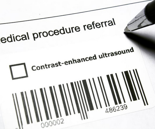
milla1cf Wed, 04/24/2024 - 19:18 April 24, 2024 — The International Contrast Ultrasound Society ( ICUS ) and Northwest Imaging Forums ( NWIF ) announced an educational partnership to help train sonographers to perform contrast-enhanced ultrasound (CEUS) examinations and administer intravenous ultrasound contrast agents.
Point of care ultrasound (POCUS) is revolutionizing the healthcare industry and changing how doctors prescribe treatment for patients. POCUS is a diagnostic tool that utilizes ultrasoundimaging to diagnose, monitor, and guide treatments for medical conditions. What Is Point of Care Ultrasound (POCUS)?
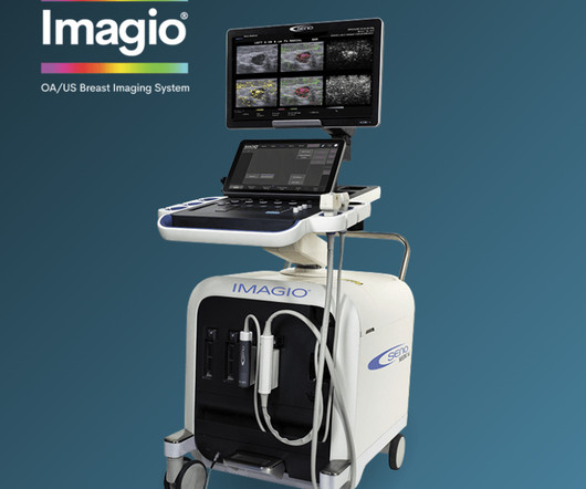
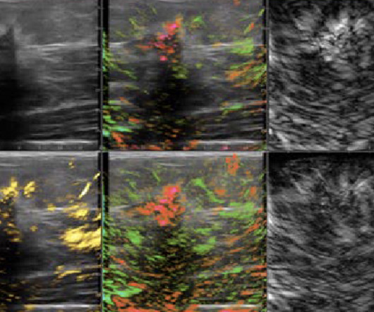
The result is a diagnosticimaging modality that delivers functional information regarding suspicious breast masses, increasing confidence regarding the need for invasive breast cancer diagnostic biopsies. Seno’s Imagio OA/US technology combines light, sound, and AI to deliver new information never before available.
Gauging public attitudes around radiology workforce shortages and shifting diagnosticimaging and ultrasound examinations and ultrasounds to AI, the Canadian Association of Radiologists (CAR) is pushing for shorter medical imaging wait times and a federal investment of over $1 billion Canadian ($721 million U.S.)
The earlier breast cancer is detected through diagnosticimaging, the better chance there is for successful treatment with surgery, radiation therapy, or chemotherapy. Consistent mammography and diagnosticimaging appointments can help stop this number from continuing to rise. this year alone. this year alone.
For dense-breasted patients requiring supplemental imaging, MRI remains a valuable option that is not limited by breast density and is shown to be more sensitive than mammography at finding breast cancer. vi Investigations continue of this newer imaging modality, which has the potential to positively benefit patients with dense breasts.
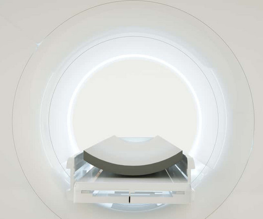
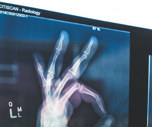
milla1cf Thu, 08/24/2023 - 16:02 August 24, 2023 — Medical imaging via X-rays , CT scans , MRIs and ultrasounds provide health-care professionals with unique perspectives and a better understanding of what’s happening inside a patient’s body. Using various forms of waves, these machines can visualize many unseen ailments and diseases.
In order to diagnose your condition, your neurologist may recommend getting an ultrasound. Also called sonography, ultrasound uses high-frequency sound waves to capture images of structures in the body. An ultrasound permits a specialist to view organs and to assess whether they are normal or if disease or injury is present.
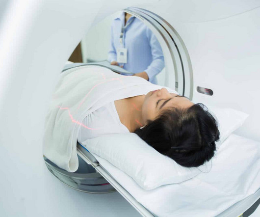
The balance of dose and image quality is even more important in pediatric medical imaging. Not only are children more radiosensitive than adults (the cancer risk per unit dose of ionizing radiation is higher), but children also have a longer expected lifetime, which puts them at greater risk of cancer following radiation exposure.(1)
Image courtesy of Canon. The company also noted that its Advanced intelligent Clear-IQ Engine (AiCE) deep learning software – which supports high-quality imaging at low radiation doses -- has been incorporated into all of its CT systems. In its booth, Canon also highlighted an automated "GPS-like" guidance tool for ultrasound.
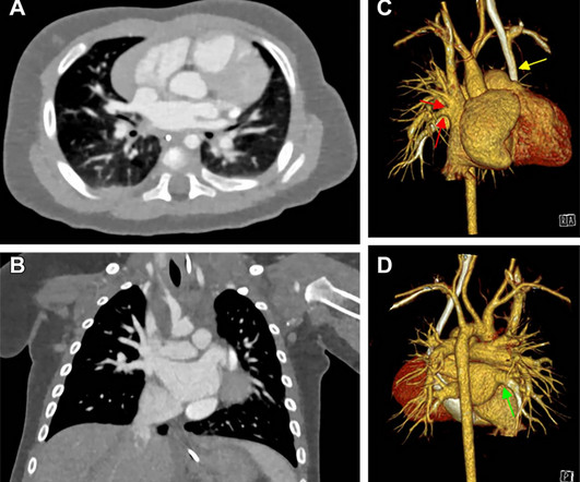
A comprehensive assessment, including ultrasound, MRI and CT exams, is typically needed to plan for surgery and to create virtual and printed 3D reconstructions of the heart. PCCT is an emerging imaging technique that counts the exact number and measures the energy of incoming x-ray photons.
You now have access to high-quality, low-cost ultrasound in King George at our newest outpatient facility. Located at 11131 Journal Pkwy, Medical Imaging of King George offers ultrasoundimaging services by appointment Monday through Friday from 8:30 a.m. Why Choose Ultrasounds at Medical Imaging of King George?
Carolina Breast Imaging Specialists, PLLC in Greenville, North Carolina intends to change that as the first radiology center in the U.S.A. to adopt the recently available Imagio Breast Imaging System. And it does this without exposing patients to potentially harmful ionizing radiation ( x-rays ) or contrast agents.
Medical imaging is a technology which is used by radiologists , particularly for diagnostic purposes. Although the word “radiology” sounds like it involves radiation, that is not always the case – for example, MRI (magnetic resonance imaging) and ultrasound do not use radiation in their medical imaging technologies.
Discuss how timely and precise imaging can significantly impact treatment outcomes and overall well-being. Multimodal Imaging Techniques in Pediatric Radiology: Explore the variety of imaging techniques employed by pediatric radiologists.
A radiology department is responsible for providing diagnostic studies and radiation therapy. Some common radiology studies include x-rays, CT scans, MRIs, mammography, and ultrasound. Because they work with radiation, there are radiation exposure risks associated with any job in radiology.
One additional consideration when imaging pediatric patients is their exposure to ionizing radiation. While MRI and Ultrasound produce NO ionizing radiation, X-Ray, CT, PET, and Nuclear Medicine studies do and therefore must be scrutinized when recommending and/or performing radiologic examinations.
Outpatient radiology centers play a crucial role in the healthcare landscape by providing convenient, efficient, and cost-effective access to diagnosticimaging services for patients across a wide range of medical conditions. These services include X-rays, ultrasounds , MRIs, CT scans, mammography, and fluoroscopy, among others.
Over the next 130 years, medical imaging has undergone many varied evolutions to become a cornerstone of modern-day medicine. Radiography has exploded into a variety of modalities and specialisms from CT to Ultrasound to MRI; all driven by research and development. All beginning with that first piece of research.
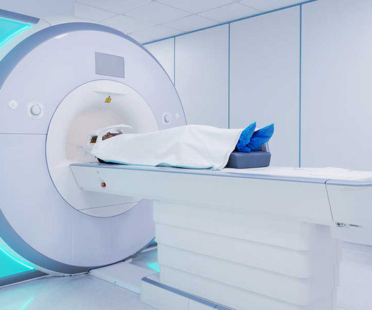
Preoperative brain imaging (usually MR imaging) is used principally for the selection of those patients with PD who are candidates for DBS intervention (bilateral GPi or STN DBS). In most cases, the presence of abnormalities on MR imaging such as severe atrophy, leukoencephalopathy, or multiple lacunae contraindicates DBS surgery.
Joint Commission on the dangers of medical radiation. Joint Commission has issued an alert on the dangers of medical radiation, sending an unmistakable signal that radiology has entered its sights in a formal way. New articles will be published each Monday until our official anniversary at RSNA 2024. Noting that the U.S.
Diagnosticimaging tests are tools used by physicians to diagnose a range of medical conditions. Each of these imaging methods uses different technologies to create real-time images and videos of the internal structures of the body. There is no need to be anxious as imaging tests are non-invasive and painless.
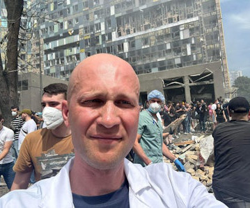
X-ray and ultrasound machines were badly damaged in a rocket attack on Ukraine's largest children's hospital on July 8, according to radiologist Stanislav Rebenkov, MD. It will take months to recover, he said. This is a very big crime," he told AuntMinnnie.com in a phone interview on July 9. There are broken windows everywhere. local time.
UEAs do not contain dye, create no known risk of kidney damage or deposit of contrast media in the brain, and do not expose patients or hospital staff to ionizing radiation. These guidelines address dilution and other evidence-based options for optimal UEA administration.
MRI Imaging of Prostate was started sometime during the mid-1980’s. Other imaging modalities that can be used in concurrence with multiparametric MRI are multiparametric ultrasound (mpUS) and nuclear imaging which further assists in accurate diagnosis and treatment guidance.
We organize all of the trending information in your field so you don't have to. Join 5,000 users and stay up to date on the latest articles your peers are reading.
You know about us, now we want to get to know you!
Let's personalize your content
Let's get even more personalized
We recognize your account from another site in our network, please click 'Send Email' below to continue with verifying your account and setting a password.


Let's personalize your content