This site uses cookies to improve your experience. To help us insure we adhere to various privacy regulations, please select your country/region of residence. If you do not select a country, we will assume you are from the United States. Select your Cookie Settings or view our Privacy Policy and Terms of Use.
Cookie Settings
Cookies and similar technologies are used on this website for proper function of the website, for tracking performance analytics and for marketing purposes. We and some of our third-party providers may use cookie data for various purposes. Please review the cookie settings below and choose your preference.
Used for the proper function of the website
Used for monitoring website traffic and interactions
Cookie Settings
Cookies and similar technologies are used on this website for proper function of the website, for tracking performance analytics and for marketing purposes. We and some of our third-party providers may use cookie data for various purposes. Please review the cookie settings below and choose your preference.
Strictly Necessary: Used for the proper function of the website
Performance/Analytics: Used for monitoring website traffic and interactions
We guarantee accuracy, clarity, and care in each scan with a staff of skilled radiologists and cutting-edge equipment. The Power of Diagnostic Imaging in Early Disease Detection Medical imaging is one of the most significant developments in medical science. What is Diagnostic Imaging? How Does Early Detection Help?
Most radiologists have heard of Moore’s Law. A close colleague observed that radiologists are affected by a type of Moore’s Law. The amount of scans a radiologist is expected to shift per day is increasing year on year. Today’s radiologist is expected to shift work at an eye-watering rate. Paul McCoubrie, MBBS.
The voters also zeroed in on the ongoing shortage of radiologists as the Biggest Threat to Radiology. He earned his medical degree at the University of Alabama School of Medicine and completed a residency in diagnostic radiology at the University of Florida. But these barriers aren't deal-breakers.
Diagnostic radiology MRI Safety A new CPT subsection has been established for reporting six new codes describing MR safety services, including implant or foreign body evaluation, safety consultation, electronics preparation, and implant positioning or immobilization. PC-1.97 $120.01 $63.72 64467 unilateral; by infusion(s) G-6.86
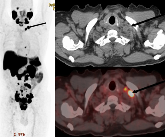
A team led by radiologists in Nairobi, Kenya, found metastases in supraclavicular (SC) lymph nodes in 8% of a large group of patients, and the group suggested adding consideration of the site during the initial diagnostic workup of patients.
Oxford Brain Diagnostics' CDM Insights software has been granted 510(k) clearance by the U.S. Healthcare practitioners can use this data to evaluate patients with neurodegenerative disorders such as Alzheimer's disease. Food and Drug Administration (FDA). The software includes new measurements of microstructure and cortical thickness.
Understanding the mechanics of flow artifacts on CT or CT angiography (CTA) and how these artifacts are created is key to better disease diagnosis, according to a review published April 25 in RadioGraphics. At first glance, flow artifacts may appear as a simple distractor to the discerning eye of a radiologist," Robb and colleagues noted.
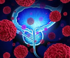
When radiologists interpretations are indeterminate, a commercially available deep learning algorithm could find clinically significant prostate cancer with improved specificity while maintaining sensitivity, suggested research published March 11 in Radiology. A radiologist also detected the lesion and assigned it PI-RADS 4 (arrow in A-C).
Diagnostic Imaging is a great tool for your medical professional to use to detect issues sooner rather than later. There are several types of diagnostic imaging available today; each one used to visualize the internal structures of the body to assist doctors in diagnosis and treating various diseases and medical conditions.
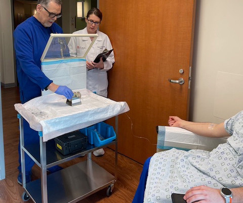
Our study showed that the availability of PET/CT prior to the [percutaneous needle lung biopsy] improves the diagnostic biopsy rates,” wrote lead author Konstantinos Stefanidis, MD, of King’s College Hospital in London, and colleagues. Patient with extensive lung and pleural disease. (2a) A team that included researchers in the U.K.,
A deep-learning model performs comparably to an abdominal radiologist when it comes to finding clinically significant prostate cancer on MRI, researchers have reported. External 0.86 External 0.86
A thyroid ultrasound imaging database could spur the development of more effective diagnostic and treatment models for related diseases, according to research published January 23 in Ultrasound in Medicine & Biology. One reason is that radiologists place markers on images to outline where the lesions are to inform other clinicians.
and radiology has learned much since then, according to experts who directly dealt with the diseases impact. While the pandemic affected medical operations across the country, the experts said that radiologists developed and honed their sense of resiliency as imaging was placed on the front lines.
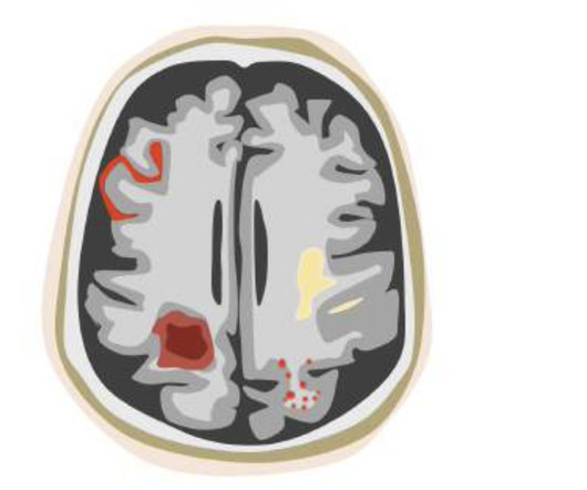
This will help radiologists and neurologists across India leverage software to improve diagnostic accuracy and patient outcomes, Cortechs.ai has developed AI software for use in assessing neurological disorders such as Alzheimers disease, epilepsy, multiple sclerosis, brain trauma, tumors, and other brain abnormalities.
Other potential uses researchers are investigating are filtering out normal chest radiographs, monitoring reading errors, and automated opportunistic screening of incidentally identified disease. These LLMs offer opportunities ranging from generating text reports from images to explaining examination results to patients."
milla1cf Tue, 09/26/2023 - 15:32 September 26, 2023 — In a study of more than 2,000 chest X-rays , radiologists outperformed AI in accurately identifying the presence and absence of three common lung diseases, according to a study published in Radiology , a journal of the Radiological Society of North America ( RSNA ). Plesner, M.D.,
A large study, needed for FDA clearance, demonstrated that the use of icobrain aria significantly increases the accuracy of ARIA assessments by radiologists and hence allows for safer use of new amyloid-beta targeting therapies for Alzheimer’s disease patients. icobrain aria was thoroughly evaluated in large reader studies.
“Skin biopsy is the gold standard for diagnosing calciphylaxis, but it can worsen lesions and confer poorer disease prognosis,” the researchers wrote, in a study published October 23 in JAAD International. The team assessed the potential diagnostic utility of bone scans in calciphylaxis based on a review of the literature.
Becoming a radiologist requires an extraordinary level of dedication, education, and training. Radiologists are medical doctors who specialize in interpreting imaging studies like X-rays, CT scans, MRIs, and ultrasounds to diagnose and guide treatment for various conditions.
If MRI prostagram should prove to be a good screening tool for prostate cancer, urologists are going to have a lot of work on their hands, and radiologists will be needed to overcome this burden, said the U.K. MRI reports should include a quality reference remark that precedes the diagnosis, explained radiologist Jelle Barentsz, MD, PhD.
Healthcare disparities continue to plague medical imaging, but there are concrete measures radiologists can take to mitigate them, according to a paper published on October 12 in RadioGraphics. Histopathologic biopsy results demonstrated invasive mammary carcinoma with metastatic disease in a right axillary lymph node.
Part of the trial's overarching work includes predicting whether complete debulking surgery could be achieved and whether or not DW-MRI could be a cost-effective method in the diagnostic work-up of advanced ovarian cancer. that every visible disease has to be removed surgically," Lahaye said. Complete debulking is very important.
The authors of a practical guide designed to help radiologists avoid malpractice claims have received one of 17 magna cum laude awards handed out by the RSNA 2023 judges. The majority of radiologists will face litigation in their careers," noted Luke Michael Wojdyla, a second-year radiology resident, and radiologist James Y.
The responses were scored as correct, incorrect, or clinically misleading by two cardiologists and one radiologist, with categorization based on majority vote. Questions ranged from those on simple diagnostic subjects (“My doctor wants to run tests to diagnose coronary artery disease. What tests will be ordered?”)
A novel machine-learning algorithm used with MRI can harmonize brain volumetric data of patients undergoing Alzheimer's disease assessment gathered from different scanners, researchers have reported. The findings were published December 18 in Radiology: Artificial Intelligence. The complete study can be found here.
Integrating AI with our advanced imaging techniques will allow us to detect subtle changes in brain activity indicative of neurodegenerative diseases at their nascent stage. The London Ontario, Canada-based company's focus on neuroimaging analysis and classification cements its position as a leader in neurological innovation.
Using AI software with brain MR imaging improves the diagnostic accuracy for the monitoring of amyloid-related imaging abnormalities (ARIA) in patients undergoing beta amyloid-directed antibody therapies for Alzheimer's disease, researchers have found. But detecting ARIA and assessing its severity can be tricky. ARIA-H detection 0.78
Chest x-rays are the go-to modality for assessing whether or not a disease requires immediate treatment. That last part can be challenging for nonradiologists who do not constantly interpret diagnostic imaging exams.
Imaging was so important [for cardiac indications], that I decided to become a radiologist," he said. He noted that in 2019, the European Society of Cardiology issued updated guidelines for diagnostic imaging of coronary artery disease (CAD), recommending noninvasive imaging (i.e., But he persevered. Just do it!"
His research interests include using structural and functional MRI -- particularly ultrahigh-field, 7-tesla MRI -- to map brain microstructure and develop neurosurgical treatment of brain tumors, epilepsy, and neurodegenerative and movement disorders such as Parkinson's disease, essential tremor, and dystonia. He served in the U.S.
Patients prefer structure and thorough details on radiology reports, according to research published October 21 in Current Problems in Diagnostic Radiology. They wrote that such tools could provide patient-friendly reports automatically while still including validated and well-thought-out educational materials created by radiologists.
The precision and accuracy of diagnostic procedures have made remarkable strides. Professional Radiology, a leading diagnostic imaging radiology center in El Paso, has been at the forefront of these innovations, offering a wide range of services to aid in the early detection and diagnosis of diseases.
Methods of detection included screening mammography in asymptomatic patients, patient or provider disease identification through self or clinical examination, and an alternative identification of disease (for example, incidental finding on CT imaging).
History of CT image recon algorithms For added historical perspective, radiologists at Stanford University, University of Wisconsin-Madison, and Leiden University Medical Center in the Netherlands early last year compiled a simple history of CT image reconstruction algorithms.
Eliot Siegel, MD; Stanislav Spiridonov, MD; Nathan Gee, MD; and Anthony Chang, PhD, are among a niche gathering of early adopters, entrepreneurial physicians, medical physicists, and investors with a sweet spot for nuclear medicine, diagnostic radiology, and radiation oncology.
Considering a patient's symptoms as noted on a short pre-MR imaging questionnaire improves radiologists' lumbar spine exam interpretation and diagnosis, researchers have found. Diagnostic certainty was higher for those MRI exam readings that included data from patients' symptom questionnaires compared with those that did not (mean, 80.4
Researchers are warning that the COVID-19 vaccine can manifest on imaging in ways that appear to be disease, according to two studies published February 24 in the American Journal of Roentgenology and Radiology. Ultrasound from diagnostic work-up performed seven days later showed no change in lymph node size. BI-RADS 3 was assigned.
The diagnosis and management of intermediate-risk and high-risk prostate cancer are increasingly informed by advances in diagnostic imaging, the authors noted. Radiologists and nuclear medicine physicians analyzed the images, with their reads ultimately compared to laboratory results from biopsies.
news media, though radiologists may not be included in such coverage, researchers have reported. However, radiologists were “infrequently” interviewed or quoted in these articles. However, radiologists were “infrequently” interviewed or quoted in these articles. The findings were published July 20 in Clinical Imaging. “It
S1-SSCH01-2 | E451A Computing thoracic bone and muscle metrics in patients with chronic obstructive pulmonary disease (COPD) improves understanding of the role of certain comorbidities in COPD disease progression, according to findings to be announced in this scientific presentation. 9:20 a.m. | 11:00 a.m. | 2:40 p.m. | 3:30 p.m. |
Lungscreen Australia has adopted Annalise.ais clinical decision-support technology to provide its radiologists with AI tools with the goal of faster, more accurate diagnostics, according to the organization.
milla1cf Wed, 08/02/2023 - 19:58 August 2, 2023 — An accepted manuscript published in the American Journal of Roentgenology (AJR) found that deploying a radiomic-based model with T2-weighted MRI data could increase diagnostic accuracy for pediatric Crohn disease (CD). and accuracy of 89.6%. and accuracy of 93.5%.
Theranostics pairs diagnostic biomarkers that can be visualized on nuclear medicine imaging with therapeutic agents that share a specific target in diseased cells or tissues. Traditional treatments didn't slow the disease in these patients, noted UT’s Vroman.
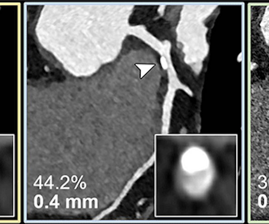
Coronary CT angiography is a first-line test in the assessment of coronary artery disease. However, its diagnostic value is limited in patients with severe calcifications, or calcium buildup in the plaque of the coronary arteries. The technology has the potential to improve patient management and reduce unnecessary interventions.
We organize all of the trending information in your field so you don't have to. Join 5,000 users and stay up to date on the latest articles your peers are reading.
You know about us, now we want to get to know you!
Let's personalize your content
Let's get even more personalized
We recognize your account from another site in our network, please click 'Send Email' below to continue with verifying your account and setting a password.


Let's personalize your content