This site uses cookies to improve your experience. To help us insure we adhere to various privacy regulations, please select your country/region of residence. If you do not select a country, we will assume you are from the United States. Select your Cookie Settings or view our Privacy Policy and Terms of Use.
Cookie Settings
Cookies and similar technologies are used on this website for proper function of the website, for tracking performance analytics and for marketing purposes. We and some of our third-party providers may use cookie data for various purposes. Please review the cookie settings below and choose your preference.
Used for the proper function of the website
Used for monitoring website traffic and interactions
Cookie Settings
Cookies and similar technologies are used on this website for proper function of the website, for tracking performance analytics and for marketing purposes. We and some of our third-party providers may use cookie data for various purposes. Please review the cookie settings below and choose your preference.
Strictly Necessary: Used for the proper function of the website
Performance/Analytics: Used for monitoring website traffic and interactions
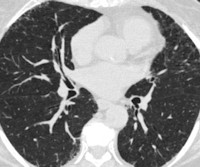
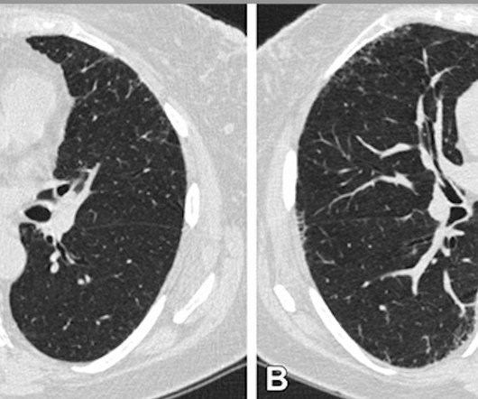
CT imaging shows that severe acute respiratory disease events can be caused by quantitative interstitial abnormalities (QIA) -- that is, small irregularities that don't necessarily meet diagnostic criteria for advanced pulmonary diseases but show up on CT exams over time, a study published April 30 in Radiology has reported.
A steady increase in the volume of coronary CT angiography (CCTA) exams is occurring, especially at mid- to high-level community hospitals across the country. The radiology department must be able to post-process the images, edit them, and make them diagnostically useful at all hours of the day and night.
Substituting less energy-consuming ultrasound for x-ray or CT reduced energy use by as much as 8% during diagnostic radiology processes and 31.2% The group developed a "green imaging review" for patients accessing the emergencyroom. for total CFCs, while demonstrating an increase in diagnostic activity of 11.8%, Masperi said.
That’s clearly the case at Massac Memorial Hospital , an award-winning critical access hospital in Metropolis, Ill., The hospital serves patients in Massac County and the surrounding areas. “Our The recent investment in a full portfolio of diagnostic imaging equipment is the latest testament to that philosophy.
At lower radiation doses, clinicians can detect and evaluate small structures precisely and with fewer artifacts, maximizing the diagnostic information and accuracy for fast decision-making and optimized imaging workflows. Photon-counting computed tomography will replace conventional CT examinations in the medium term.
Diagnostic errors, including missed or delayed diagnosis of vascular events, account for the single largest source of medical harm and death to patients each year. AI’s Role in Addressing Diagnostic Challenges Imaging remains central to the diagnosis and management of all stroke types.
Emergencyrooms (ERs) play a critical and indispensable role in the healthcare system, serving as the front line of medical care for individuals experiencing urgent and life-threatening situations. During the summer months in the United States, emergencyrooms tend to see an increase in patient visits due to various reasons.
Quantitative interstitial abnormalities (QIA) are subtle abnormalities on chest CTs that do not meet the diagnostic criteria for advanced pulmonary diseases but are nonetheless associated with decreased lung function and capacity, increased respiratory symptoms and death. “QIA
Tailored for use in the incidental adult population, this innovative device is a game-changer in diagnostic technology. The performance of various readers, including radiologists, pulmonologists, and emergencyroom physicians, showed improvement. stands at the forefront of diagnostic advancement. Diagnostics 2022).
The common adage of a bustling and stressful emergencyroom is resoundingly accurate. Communication between medical providers, specifically emergency physicians and Radiologists , drive clinical decisions that facilitate the coordination of the patient journey from arrival to diagnostic imaging to eventual admission or discharge.
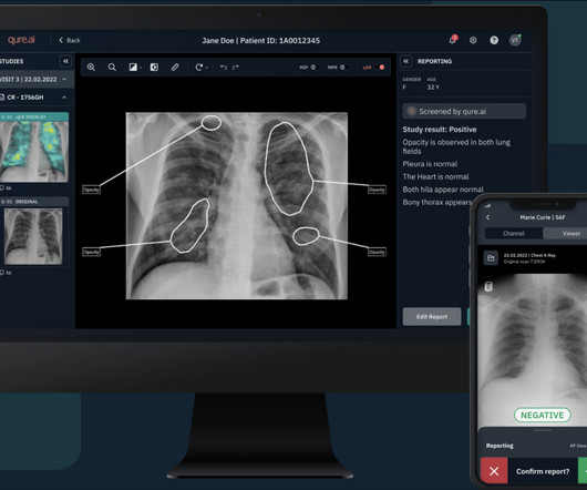
The findings boost AI-driven medical diagnostics and bring health care professionals closer to being able to quickly diagnose patients with COVID-19 and other pulmonary diseases with algorithms that comb through ultrasound images to identify signs of disease.
The common adage of a bustling and stressful emergencyroom is resoundingly accurate. Communication between medical providers, specifically emergency physicians and Radiologists , drive clinical decisions that facilitate the coordination of the patient journey from arrival to diagnostic imaging to eventual admission or discharge.
A good portion of inpatient hospital admissions are funneled through the ED. Several factors contribute to delays and overcrowding in the emergency department, such as: The increasing demand for emergency services, driven by factors such as population growth, aging demographics and limited access to primary care services.
A radiology department is responsible for providing diagnostic studies and radiation therapy. Radiologic techs take diagnostic images of patients and ensure that the images are clear enough for radiologists to interpret. Trauma patients are common, some coming directly from the emergencyroom.
Swift Diagnosis in Emergency Settings: In the bustling emergencyrooms of urban hospitals and the remote clinics in rural areas, teleradiology has emerged as a lifesaver. Rural healthcare centers, once limited in diagnostic capabilities, now connect seamlessly with urban-based radiologists.
Our radiologist was quick to respond to review the x-ray and advised the patient to go to the hospital. Emergency Medical Services (EMS) was notified immediately and an ambulance arrived to take this gentleman, who lives alone and has no family support, to the EmergencyRoom. Kudos to our team at Homosassa Open MRI.
5] [6] As the entry point to diagnostic imaging in the emergencyroom, inpatient bedside imaging and the intensive care unit (ICU), GE HealthCare continues to reinvent mobile X-ray to be one of the most intuitive and technologically powerful imaging tools available to help clinicians respond fast without compromising diagnostic precision.
Clinical Case: Getting the Care You Need Mark is a 55-year-old male with a history of diabetes, hypertension and hyperlipidemia who comes to the emergencyroom with a chief complaint of shortness of breath. He informs the nurse that he has been smoking since the age of 25.
However, evaluating and managing patients with acute alcohol intoxication in the emergency department can be challenging. Patients may be agitated or altered, hindering their initial evaluation and diagnostic workup. Annals of Emergency Medicine. 2018 Mar;71(3):279-288. PMID: 28844504 Suokas J, Lonnqvist J. Acta Psychiatr Scand.
It should be a very simple problem for professionals who rely on diagnostic tests for a living: in a sample of 10,000 people, 10 have the disease and get a true positive result; 5 percent, or 500, will get a false positive; out of 510 people who test positive, only 10, or 1.96 The most common answer was 95 percent.
We organize all of the trending information in your field so you don't have to. Join 5,000 users and stay up to date on the latest articles your peers are reading.
You know about us, now we want to get to know you!
Let's personalize your content
Let's get even more personalized
We recognize your account from another site in our network, please click 'Send Email' below to continue with verifying your account and setting a password.


Let's personalize your content