This site uses cookies to improve your experience. To help us insure we adhere to various privacy regulations, please select your country/region of residence. If you do not select a country, we will assume you are from the United States. Select your Cookie Settings or view our Privacy Policy and Terms of Use.
Cookie Settings
Cookies and similar technologies are used on this website for proper function of the website, for tracking performance analytics and for marketing purposes. We and some of our third-party providers may use cookie data for various purposes. Please review the cookie settings below and choose your preference.
Used for the proper function of the website
Used for monitoring website traffic and interactions
Cookie Settings
Cookies and similar technologies are used on this website for proper function of the website, for tracking performance analytics and for marketing purposes. We and some of our third-party providers may use cookie data for various purposes. Please review the cookie settings below and choose your preference.
Strictly Necessary: Used for the proper function of the website
Performance/Analytics: Used for monitoring website traffic and interactions
A team of researchers at Boston Children’s Hospital has developed an age-specific dose catalog for estimating radiation exposure to children from diagnostic and interventional radiology fluoroscopy procedures. They analyzed metrics to estimate age-specific effective dose per IR procedure type and diagnosticfluoroscopy exam.
To cover a CT and MR "list" was a luxury; it was to escape the murderous fluoroscopy rooms, intravenous urography (IVU) lists, and indeterminable piles of plain films Then the scanners got fast. So as to report more CT and MRI, radiologists stopped doing hands-on ultrasound and fluoroscopy. 20s rotation, 40s reconstruction.
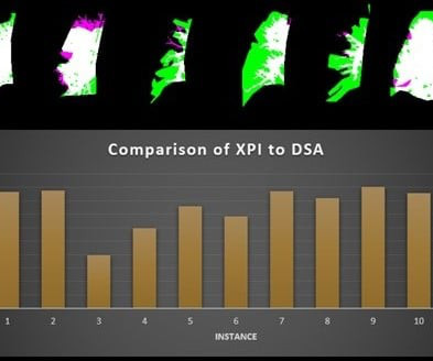
A fluoroscopy method incorporating a noncontrast x-ray pulsatility index (XPI) can improve clinical efficiency as a screening or diagnostic test for suspected chronic thromboembolic pulmonary hypertension, according to research presented at the American Roentgen Ray Society (ARRS) annual meeting.
In the realm of regenerative medicine, the integration of advanced imaging technologies like digital X-ray and fluoroscopy has become paramount. These tools not only enhance diagnostic accuracy but also revolutionize the administration and assessment of regenerative treatments.
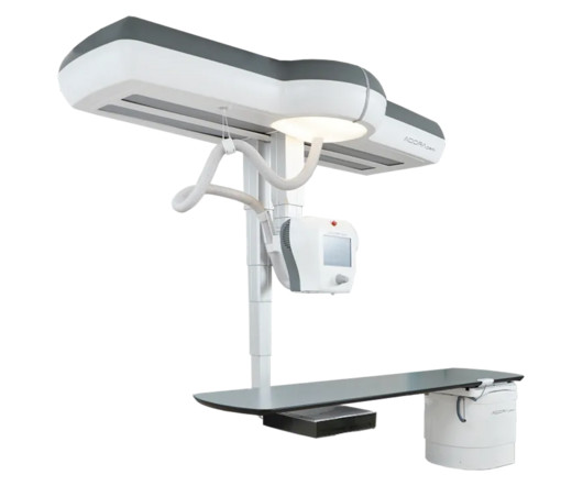
Enabling radiographic and low-dose fluoroscopy exams in the same room, the Adora DRFi system emphasizes a variety of advances for flexibility with patient positioning.
In addition, they computed the out-of-network rate by imaging modality, with claims categorized as CT, MR, nuclear medicine (NM), ultrasound, radiography or fluoroscopy (XR), and interventional procedures. For example, among diagnostic imaging, NM had the largest decline, from 14.5% In total, they identified 80.4 in 2007 to 1.1%
The group used the National Cancer Institute dosimetry system for radiography and fluoroscopy (NCIRF) to estimate absolute organ-specific radiation doses in 10 cardiac catheterizations and 20 diagnostic fluoroscopic procedures in a cohort of neonates with cyanotic CHD who required early intervention. Louis, and colleagues.
to identify noninvasive diagnostic imaging services in this data. Ultrasound 24% 28% X-ray/fluoroscopy 56.2% Ultrasound 24% 28% X-ray/fluoroscopy 56.2% million individuals; they used the Neiman Imaging Types of Services (NITOS), version 4.0 The changes varied by modality, according to the researchers. Nuclear medicine 2.7%
To address this knowledge gap, Christensen and colleagues conducted a study that assessed associations between diagnostic imaging use with the state-level professional payment MMRR, computing these ratios by imaging modality. X-ray or fluoroscopy 0.82 Nuclear medicine 0.76 Ultrasound 0.85 higher for MR, 21.4% higher for x-ray.
Diagnostic radiology Coronary Fractional Flow Reserve (FFR) with CT : New Category I code 75580 will replace Category III codes 0501T, 0502T, 0503T, and 0504T to describe noninvasive estimated coronary FFR derived from augmentative AI software analysis of coronary CT angiography (CCTA) data.
Thiel and colleagues performed an LCA in April 2023 of the diagnostic radiology practice of Vanderbilt University Medical Center in Nashville, TN. "LCAs establish baseline data on carbon emissions and environmental waste and help analyze the impact of interventions on carbon footprints."
The study, published in the journal Diagnostics, highlights the clinical value of DDR through its unique ability to evaluate diaphragm movement in real time and integrate dynamic functional information with static anatomical data to provide a quantitative assessment of diaphragmatic movement, including excursion and speed.
Onsite procedures include general fluoroscopy, minor ultrasound, paracentesis, and thoracentesis. Onsite procedures include general fluoroscopy, minor ultrasound, paracentesis and thoracentesis. Onsite procedures include general fluoroscopy, minor ultrasound, paracentesis and thoracentesis. No neuro or MSK. No neuro or MSK.
milla1cf Thu, 11/23/2023 - 06:00 November 23, 2023 — Fujifilm Healthcare Americas Corporation, a leading provider of diagnostic and enterprise imaging solutions, is unveiling several new medical systems at the 2023 Radiological Society of North America ( RSNA ) annual meeting, booth #1929, held November 26 – 30 at McCormick Place in Chicago.
Our hope is that we'll be able to determine how to use this as a diagnostic tool in actual practice,” he said. He added that by the age of 50 or 60, many or most people have already had a CT scan for diagnostic purposes. “We We can first take advantage of all those scans,” he said.
Offering ease of mobility and self-driving capabilities, the Ciartic Move C-arm device reportedly reduces the stress and potential for error associated with manual repositioning during intraoperative imaging with computed tomography and fluoroscopy.
Additionally,” said Matthew Smith , MD, from Vanderbilt University Medical Center in Nashville, TN, “this easy-to-implement method can be performed by an x-ray technologist in an outpatient setting,” Smith et al. enrolled volunteers suspected of chronic thromboembolic pulmonary hypertension (CTEPH) based on pulmonary scintigraphy and/or CTA.
Central Vein Sign in Multiple Sclerosis: A Comparison Study of the Diagnostic Performance of 3T versus 7T MRI. Brain fog in long COVID: A glutamatergic hypothesis with astrocyte dysfunction accounting for brain PET glucose hypometabolism. Horowitz, et al, Medical Hypotheses , October 13, 2023. To learn more about this paper, click here.
With Caption Guidance, even healthcare providers without specialized training or experience can perform cardiac ultrasound exams and acquire diagnostic-quality cardiac images, according to the vendor.
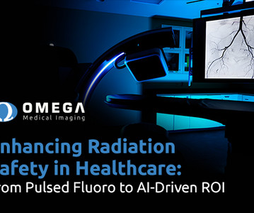
Initially, pulsed fluoroscopy marked a huge shift from continuous fluoroscopy by considerably reducing radiation exposure while preserving diagnostic accuracy.
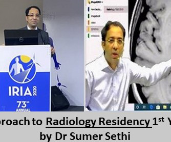
As far as PG is concerned, the most common practice is to teach Diagnostic radiology. Many are of the opinion that diagnostics is mastered mostly by practical work. It is crucial to regularly observe various reports like MRI, CT scan, X-Ray, Doppler, Fluoroscopy etc. You will find intervention radiology in a handful of places.
Teleradiology-India Introduction: “Capturing the Invisible” takes you on a captivating journey into the world of X-rays, unveiling their impactful role in diagnostics. How X-rays are generated, interact with the human body, and create diagnostic images.
This blog explores the critical role of radiology in on-the-field diagnostics for sports-related injuries. C-Arm Fluoroscopy: Real-Time Imaging in Interventional Procedures: Highlight the use of C-arm fluoroscopy for real-time imaging during interventional procedures.
Unlike fluoroscopy, DDR is a series of individual digital X-ray images acquired at high speed and low doses, allowing the visualization of anatomy in motion. Greg Miner, Appleton Area Health Chief Executive Officer, says, “To bring this type of diagnostic care to Appleton is truly remarkable.
milla1cf Fri, 06/21/2024 - 20:24 June 21, 2024 — GE HealthCare , a leading global medical technology, pharmaceutical diagnostics, and digital solutions innovator, and MediView XR Inc.,
To meet that goal, we had to get the best possible equipment and that’s why we turned to Fujifilm Healthcare Americas Corporation for the very latest in diagnostic imaging systems.” The recent investment in a full portfolio of diagnostic imaging equipment is the latest testament to that philosophy.
CT LVAS, designed for use with computed tomography scans, joins 4D Medical's XV LVAS imaging software cleared for use with fluoroscopy in the United States. As we head to RSNA, I am thrilled that reimbursement has been approved for our XV LVAS product designed for fluoroscopy, and to also share FDA clearance for CT LVAS in the U.S.
A study published in Current Problems in Diagnostic Radiology examines the increasing trend of NPPs taking on imaging interpretation responsibilities. Most NPP interpretations were for radiography/fluoroscopy (53.3%) or ultrasound (26.1%). NPP-billed interpretation claims increased from 2.6% in 2016 to 3.3% growth during this period.
In this exploration, we will weave together the history, science, and practical applications of X-ray imaging, providing a holistic understanding of this invaluable diagnostic tool. How X-rays are generated, interact with human tissue, and create diagnostic images. Real-life case studies illustrating the diagnostic power of X-rays.
Benefits of Teleradiology to Telehealth Introduction: “Behind the Beams” is an intriguing journey that delves into the intricate world of X-ray technology, uncovering the perfect fusion of art and science that powers this essential diagnostic tool. How X-rays are generated, interact with matter, and produce diagnostic images.
How X-rays are generated, interact with the human body, and create diagnostic images. Chapter 4: Beyond Radiography: Advanced X-ray Modalities An examination of advanced X-ray modalities, including fluoroscopy, computed tomography (CT), and mammography.
From its discovery by Wilhelm Roentgen to modern applications, we will illuminate the shadows to provide a deep understanding of this essential diagnostic tool. The historical context of Wilhelm Roentgen’s discovery and the birth of this revolutionary diagnostic tool. How each modality is used for different clinical purposes.
Imaging technologies, including ultrasound, computed tomography (CT) and fluoroscopy give this procedure a greater degree of accuracy, identifying the precise point of delivery. This pattern further reduces the number of NSAID complications a patient may experience.
This journey takes us from the early days of X-ray discovery by Wilhelm Roentgen to the cutting-edge digital and computational innovations that shape the modern landscape of diagnostic imaging. How technological advancements have improved image quality, reduced radiation exposure, and expanded diagnostic capabilities.
Outpatient radiology centers play a crucial role in the healthcare landscape by providing convenient, efficient, and cost-effective access to diagnostic imaging services for patients across a wide range of medical conditions. These services include X-rays, ultrasounds , MRIs, CT scans, mammography, and fluoroscopy, among others.
While we have continued to support the Hospital throughout the crisis, doctors* are now able to schedule both diagnostic and screening exams in North Stafford, Fredericksburg, Lee’s Hill, and King George. Patients may also schedule mammograms directly at our facilities in these same locations.
The procedure first begins with gaining femoral/jugular vein access and inserting a stiff guidewire into the right atrium, confirmed with fluoroscopy [5,6]. 2015.11.047 Deven Champaneri is a medical student at Edward Via College Osteopathic Medicine (VCOM) – Carolinas and plans to pursue residency in diagnostic radiology.
The Radiology Experience Tour expanded this year to include a portfolio of Philips imaging solutions including Magnetic Resonance, Radiology and Fluoroscopy, Ultrasound, Radiology Operations Command Center (ROCC), PACS, C-Arms and Ambient Experience. The objectives behind the radiology experience tour are twofold.
The agreement includes hundreds of new systems, including AI-enabled technologies , across nuclear medicine , X-Ray , vascular and cardiovascular ultrasound , regional CT , fluoroscopy, surgery, and bone densitometry for which GE HealthCare will be the sole provider.
We organize all of the trending information in your field so you don't have to. Join 5,000 users and stay up to date on the latest articles your peers are reading.
You know about us, now we want to get to know you!
Let's personalize your content
Let's get even more personalized
We recognize your account from another site in our network, please click 'Send Email' below to continue with verifying your account and setting a password.


Let's personalize your content