This site uses cookies to improve your experience. To help us insure we adhere to various privacy regulations, please select your country/region of residence. If you do not select a country, we will assume you are from the United States. Select your Cookie Settings or view our Privacy Policy and Terms of Use.
Cookie Settings
Cookies and similar technologies are used on this website for proper function of the website, for tracking performance analytics and for marketing purposes. We and some of our third-party providers may use cookie data for various purposes. Please review the cookie settings below and choose your preference.
Used for the proper function of the website
Used for monitoring website traffic and interactions
Cookie Settings
Cookies and similar technologies are used on this website for proper function of the website, for tracking performance analytics and for marketing purposes. We and some of our third-party providers may use cookie data for various purposes. Please review the cookie settings below and choose your preference.
Strictly Necessary: Used for the proper function of the website
Performance/Analytics: Used for monitoring website traffic and interactions
"Practical AI implementation will require objective onsite performance evaluation, institutional information technology infrastructure integration, and postdeployment monitoring," wrote a team led by Eui Jin Hwang, MD, PhD, of Seoul National University Hospital in South Korea. van Beek, MD, of the University of Edinburgh in the U.K.
Some of my radiological heroes would report a staggering 30,000 to 40,000 radiographs a year. Some even [startled gasp] gave up reporting plain radiographs. Many diagnostic radiologists became pure CT and MRI specialists. But the pressure kept building, and the number of CT and MRI scans grew by 20% annually in my hospital.
He earned his medical degree at the University of Alabama School of Medicine and completed a residency in diagnostic radiology at the University of Florida. I'm a radiographer,' " Stewart recalled. A recent trip to Korle-Bu Teaching Hospital in Ghana has Beyder thinking about what the future holds for the international cancer center.
The finding is from a study conducted by a group at the University Hospital Jena in Eisenberg, Germany, that explored whether hip x-rays could be reliable indicators for bone mineral density (BMD) in male and female patients prior to THA procedures. “An
He earned his medical degree at the University of Alabama School of Medicine and completed a residency in diagnostic radiology at the University of Florida. An assistant professor of radiology and radiological science, Francis Deng, MD, is the Johns Hopkins radiology department's co-director of medical student diagnostic radiology electives.
tesla MRI AI body composition analysis Cardiac PET Cryo/thermoablation CT colonography Genicular artery embolization Hyperpolarized xenon-129 MRI PET/MRI Photon-counting CT Radiomics Theranostics Whole-body MRI screening Image of the Year 3D PET/MR image.
“This method plays an important role in overcoming the barriers to its clinical implementation, including the need for trained experts and the time-consuming process of manual contouring,” noted lead author Noriaki Wada, MD, of Brigham and Women’s Hospital in Boston. The subject is a 77-year-old male with VC of 1.68 L and FEV1 of 0.68
In an open forum, Yi Xiang Tay, of Singapore University Hospital's radiography and diagnostic imaging department, shared his team's research. It was the endnote of a series of sessions focused on optimizing radiology services.
Substituting less energy-consuming ultrasound for x-ray or CT reduced energy use by as much as 8% during diagnostic radiology processes and 31.2% for total CFCs, while demonstrating an increase in diagnostic activity of 11.8%, Masperi said. Masperi and colleagues conducted the study between January 2023 and June 2023.
A group at Mount Sinai Hospital developed a “pipeline” of convolutional neural networks (CNNs) to analyze lung areas in DDR image sequences from patients. Common diagnostic tests for pulmonary disorders include chest x-rays and pulmonary function tests (PFTs). a) Raw example of a dynamic digital radiograph. (b)
This competition demonstrated the value of AI in detecting and localizing many pathologies in chest radiographs by simulating the real work situations of radiologists,” the group wrote. The performance of all radiologists with and without AI assistance showed that AI improved the diagnostic accuracy of the doctors,” the group wrote.
Reading Time: 10 minutes read By Henry Williams, Carestream Area Vice President, Sales Western Nowadays, with hospital budgetary restrictions at the forefront of the purchasing decision making process, it seems like the X-Ray market, like everything else, is not immune to the current state of the economy. Who is Making the Purchases?
Whenever bilateral standing radiographs would have been needed, a WBCT was performed instead. This was the case for diagnostic purposes but also for follow-up imaging at 3, 6 and 12 months after re-alignment procedures, fusions or arthroplasties. I use WBCT every day.
11, 2025 Harrison.ai, a developer of AI-powered medical diagnostic support and workflow solutions, has announced the accelerated expansion of its operations into the United States a move supported by US$112 million of Series C funding. Radiologists using Harrison.ai's technology have seen an over 45% increase in diagnostic accuracy1.
Our hope is that we'll be able to determine how to use this as a diagnostic tool in actual practice,” he said. He added that by the age of 50 or 60, many or most people have already had a CT scan for diagnostic purposes. “We Maryland also has an all-payer system under which all payers pay the same amount for hospital-based services."
The authors note that ionizing radiation is the basis for the production of diagnostic X-rays, however it has long been proven to increase the risk of cancer. 26 –29, 2023 in North Hall, Level 3, booth #7913.
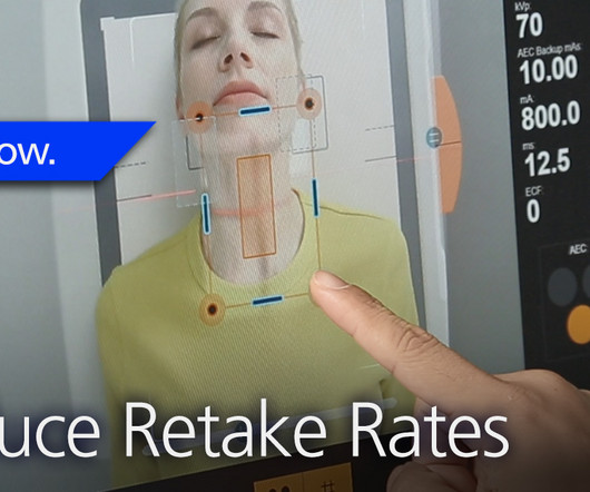
Repeating imaging exams increases the workload of your radiographers who are already stretched too thin; increases the exposure of the affected patients; and contributes to patients’ reduced confidence and satisfaction with your imaging department. The Audio Assist makes it easier for radiographers to hear the patients.
Reveal’s ability to simultaneously acquire conventional and dual-energy images with a single exposure at the bedside improves hospital and patient outcomes and protects revenue by reducing outflows. Furthermore, reimbursement for ICU patients is capitated so an unnecessary CT scan increases cost of care and the financial burden for hospitals.
Positions (On-site): Body (100% Body) – Regions Hospital Mix of shifts worked on-site Mixture of hospital, outpatient, and remote Interpret MRI, CT, U/S, and radiographs After-hours coverage provided internally by the emergency radiology section No neuro or MSK Body/Mammo – Western Wisconsin 45-minute drive from the Twin Cities.
Key Points: Currently plain radiographs are the standard method in diagnosing syndesmotic ankle injuries even though the distal tibiofibular joint cannot be assessed due to superposition of the osseous structures in the foot. Dr. Peiffer et.
The enduring shortage is affecting staffing for radiographers and radiologists; and all imaging modalities. Marion Anderson, CRA, Diagnostic Imaging Manager at Karmanos Cancer Center in Detroit, Michigan reports that her facility is down two-thirds of staff for CT and mammography exams. Bureau of Labor Statistics.
In addition, an in-person demonstration called Radiology Reimagined: AI, innovation, and interoperability in practice is designed to showcase new technologies and communications standards needed to integrate AI into the diagnostic workflow, according to the RSNA. 10:50 a.m. | 2:00 p.m. |
” The question was initially puzzling, as it seemed to have an obvious answer… but do we always inform patients appropriately of decisions around diagnostic imaging? Radiologists and radiographers undertaking the imaging have most likely never seen the patient before. RadioGraphics. Carlsson, S. 22(21-22):3225-3234.
In the third blog of her series on AI and the radiographer, Shamie Kumar explores the impact on the radiographer when AI is integrated within an imaging modality. The question to explore in this blog is when AI is integrated within an imaging modality itself and how that may impact a radiographer.
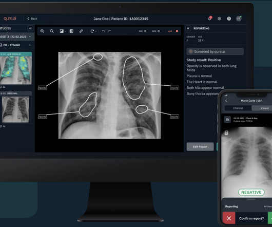
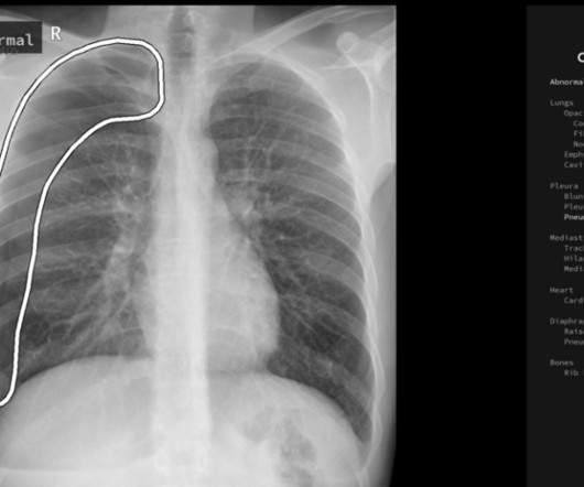
Tailored for use in the incidental adult population, this innovative device is a game-changer in diagnostic technology. It can also serve as a crucial second reader for physicians, assisting in the review of frontal (AP/PA) chest radiographs of adults acquired on digital radiographic systems. Diagnostics 2022).
But how will AI in the workplace affect the radiographer and how does it differ from the red dot system radiographers are so familiar with? The Red Dot System Often one of the first courses a newly qualified radiographer attends is the red dot course. What does AI do that a radiographer doesn’t already?
Kim Mason Kim Mason, an Audit and Research Radiographer for Mid Yorkshire Teaching Hospitals Trust, talks about their role as well as the value of radiographer engagement in research activities and how to get involved. So, what is an Audit and Research Radiographer? That is where I come in. How can I get involved?
This blog post explores the indispensable role of PACS in hospitals, highlighting their evolution, impact on healthcare workflows, and the continuous advancements that contribute to enhanced patient care. Discuss the reduction in time delays, elimination of physical storage constraints, and the acceleration of the diagnostic process.
With advance planning and an emphasis on learning, your facility can perform imaging studies with the same standard of care as a large children’s hospital. With advance planning and an emphasis on learning, your facility can perform imaging studies with the same standard of care as a large children’s hospital.
Powered by Eclipse, ImageView utilizes AI and Carestream’s proprietary algorithms to provide our most advanced image processing capabilities for superb image quality and diagnostic confidence. Physicians have better diagnostic confidence when they can view radiographs in the manner most suitable to their preferences.
milla1cf Thu, 11/23/2023 - 06:00 November 23, 2023 — Fujifilm Healthcare Americas Corporation, a leading provider of diagnostic and enterprise imaging solutions, is unveiling several new medical systems at the 2023 Radiological Society of North America ( RSNA ) annual meeting, booth #1929, held November 26 – 30 at McCormick Place in Chicago.
Figure 2 A: AP view radiograph of right forearm. B: Lateral radiograph view of right forearm. An angulated fracture of the distal midshaft radius is also visualized, but there is also bowing of the ulna that is more appreciated on the lateral radiograph view. 8 Year Old Male With Trauma Due To A Fall. Xray of the Week Figure 1.
Further insight into the Radiology departments of Government hospitals reveals that 56% of centers function without Heads of Department and 45% of the centers are understaffed. The maximum number of radiographs reported were Chest radiographs(41.7%), followed by extremity radiographs(32.1%) for trauma.
Knee osteoarthritis (OA) clinical trials results show that weight bearing CT (WBCT) imaging may offer new insights into OA pathology beyond what plain radiographs and MRI can provide. Tom Turmezei, MPhil, MA, BMBCh, PhD, FRCR, a consultant radiologist at Norfolk and Norwich University Hospitals NHS Foundation Trust in Norfolk, UK.
Secondary Safety Outcomes: The frequency of side effects related to treatment, anxiety, and depression was evaluated with the Hospital Anxiety and Depression Scale at 1 month and 3 months All-cause mortality RESULTS 50 patients were enrolled in the trial Trial enrollment was discontinued due to the COVID-19 pandemic and the cessation of funding.
A) AP radiograph of Lisfranc Fracture Dislocation demonstrates the circled “fleck sign” or Lisfranc ligament avulsion fracture fragment. (B) C) The lateral radiograph notes with a circle, the dorsal sub dislocation of the metatarsal base. Radiographs should be repeated after two weeks to ensure surgery is unnecessary.
Today, Radiology is not only aiding the diagnostic skills of the physician but also used for quantitative measurement of anatomic and functional processes, image-guided invasive interventional procedures, and an assessment of functional / performance of the organs.
Adrian Brady, presidente de la Sociedad Europea de Radiología y radiólogo consultor del Hospital de la Universidad de Mercy en Cork, Irlanda. Es director médico del Hereditary Haemorrhagic Telangiectasia (HHT) (con sede en el Hospital de la Universidad de Mercy). Radiographics (2015);35:1668-1676.
Coronavirus imaging from hospitals all over the world was collated to provide real-time COVID reporting best practice as the world started to understand the virus more. In addition, top thoracic specialist radiologists from Europe who had already experienced COVID radiology were called to report cases for NHS hospitals.
CT is the most commonly used diagnostic modality in lens dislocation and can clearly show lens displacement [4]. Radiographics. A study of 166 hospitalized cases. He plans on pursuing a residency in Diagnostic Radiology. Initial symptoms of lens dislocation include decreased visual acuity and diplopia. 1985;62(5):352-356.
These applications, cited by Jesbon, include diagnostic functions such as detecting pneumonia on chest radiographs or grading liver tumors, as well as repetitive tasks like breast or lung nodule detection.
Frontal abdomen radiograph demonstrates foreign body consistent with capsule endoscopy device (pill cam) in descending colon. 1 ] This diagnostic procedure involves swallowing a pill-sized camera that records thousands of images of the alimentary canal including the small intestine, an area difficult to examine via traditional endoscopy.
According to a paper published in RadioGraphics , deep learning has enhanced the accuracy and efficiency of medical image interpretation, aiding radiologists in diagnosing conditions such as breast cancer, brain tumors, interstitial lung disease and intracranial hemorrhages. One field that has seen substantial benefits is radiology.
I believe that the first main difference seen in the radiologists role in 2040 will be the change that artificial intelligence (AI) will have made on the diagnostic role that radiologists currently perform. It is likely that they will lead clinic with patients to discuss their imaging, as already happens in some hospitals.
We organize all of the trending information in your field so you don't have to. Join 5,000 users and stay up to date on the latest articles your peers are reading.
You know about us, now we want to get to know you!
Let's personalize your content
Let's get even more personalized
We recognize your account from another site in our network, please click 'Send Email' below to continue with verifying your account and setting a password.


Let's personalize your content