This site uses cookies to improve your experience. To help us insure we adhere to various privacy regulations, please select your country/region of residence. If you do not select a country, we will assume you are from the United States. Select your Cookie Settings or view our Privacy Policy and Terms of Use.
Cookie Settings
Cookies and similar technologies are used on this website for proper function of the website, for tracking performance analytics and for marketing purposes. We and some of our third-party providers may use cookie data for various purposes. Please review the cookie settings below and choose your preference.
Used for the proper function of the website
Used for monitoring website traffic and interactions
Cookie Settings
Cookies and similar technologies are used on this website for proper function of the website, for tracking performance analytics and for marketing purposes. We and some of our third-party providers may use cookie data for various purposes. Please review the cookie settings below and choose your preference.
Strictly Necessary: Used for the proper function of the website
Performance/Analytics: Used for monitoring website traffic and interactions
S4-SSPH02-3 | Room S404 In this session on physics in radiography, a study suggests portable x-ray systems outfitted with digital autoexposure control (DAEC) systems enhance image quality and reduce radiation doses. Chest x-ray AI evaluated in clinical routine at a Norwegian hospital Monday, December 2 | 1:30 p.m.-1:40 1:30 p.m. |
He earned his medical degree at the University of Alabama School of Medicine and completed a residency in diagnostic radiology at the University of Florida. An assistant professor of radiology and radiological science, Francis Deng, MD, is the Johns Hopkins radiology department's co-director of medical student diagnostic radiology electives.
Israel-based Nano-X Imaging (Nanox) will conduct a clinical study evaluating its Nanox.ARC 3D imaging system in a clinical outpatient setting at Clalit Health Services-owned Beilinson Hospital, part of Rabin Medical Center.
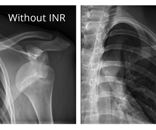
The company will showcase the clinical analysis of Canon’s Intelligent Noise Reduction (Intelligent NR) that provides superior image quality while lowering radiation dosing in pediatric digital radiography at the Radiological Society of North America Annual Meeting 2023 (RSNA) , McCormick Place Convention Center, Chicago, IL Nov.
In an open forum, Yi Xiang Tay, of Singapore University Hospital'sradiography and diagnostic imaging department, shared his team's research. It was the endnote of a series of sessions focused on optimizing radiology services.
tesla MRI AI body composition analysis Cardiac PET Cryo/thermoablation CT colonography Genicular artery embolization Hyperpolarized xenon-129 MRI PET/MRI Photon-counting CT Radiomics Theranostics Whole-body MRI screening Image of the Year 3D PET/MR image.
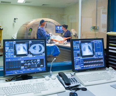
Diagnostic imaging is an important tool used every day in healthcare to assist doctors in making the most informed decisions for their patients. Over the years, advancements in diagnostic imaging have greatly increased patients’ overall care, quality of life, and outcome when diagnosed with certain conditions.
Chest dynamic digital radiography (DDR) may have received a boost toward clinical use in patients with lung disorders, with researchers developing AI to perform time-consuming analysis involved in the technology, according to researchers in New York City. The study was published March 29 in Chest Pulmonary.
Introduction : In the past decade, veterinary medicine has witnessed a transformative shift with the adoption of digital radiography systems in place of traditional film-based methods. This transition promises a myriad of benefits, ranging from improved image quality and diagnostic accuracy to enhanced workflow efficiency.
That’s clearly the case at Massac Memorial Hospital , an award-winning critical access hospital in Metropolis, Ill., The hospital serves patients in Massac County and the surrounding areas. “Our The recent investment in a full portfolio of diagnostic imaging equipment is the latest testament to that philosophy.
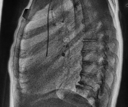
Dynamic digital radiography (DDR) has shown for the first time that it can be used to automatically capture lung signal changes during forced breathing in patients with chronic obstructive pulmonary disease (COPD), according to a recent study. and Japanese developers wrote.
Reading Time: 10 minutes read By Henry Williams, Carestream Area Vice President, Sales Western Nowadays, with hospital budgetary restrictions at the forefront of the purchasing decision making process, it seems like the X-Ray market, like everything else, is not immune to the current state of the economy.
11, 2025 Harrison.ai, a developer of AI-powered medical diagnostic support and workflow solutions, has announced the accelerated expansion of its operations into the United States a move supported by US$112 million of Series C funding. Radiologists using Harrison.ai's technology have seen an over 45% increase in diagnostic accuracy1.
announced significant expansion of X-Ray systems with Dynamic Digital Radiography (DDR) at multiple healthcare institutions across the US, including at Appleton Area Health (Appleton, Minn.), Appleton Area Health is a 15-bed critical access hospital serving a rural, mainly geriatric community that is part of a five-hospital health network.
Kim Mason Kim Mason, an Audit and Research Radiographer for Mid Yorkshire Teaching Hospitals Trust, talks about their role as well as the value of radiographer engagement in research activities and how to get involved. I’m also passionate about education, research, and (of course) radiography. That is where I come in.
Our hope is that we'll be able to determine how to use this as a diagnostic tool in actual practice,” he said. He added that by the age of 50 or 60, many or most people have already had a CT scan for diagnostic purposes. “We Maryland also has an all-payer system under which all payers pay the same amount for hospital-based services."
This was the case for diagnostic purposes but also for follow-up imaging at 3, 6 and 12 months after re-alignment procedures, fusions or arthroplasties. Cone beam CT, be it weight bearing or not, is equivalent to 3D radiography and that is where most of the healthcare benefits are for patients. I use WBCT every day.
Every November, Radiologic Technology Week celebrates the vital role radiologic technologists play in patient care and diagnostics. The Current Demand for RTs With a growing aging population, hospitals and diagnostic labs have faced shortages of skilled RTs. For instance, radiography reported an 18.1% vacancy rate.
Reveal’s ability to simultaneously acquire conventional and dual-energy images with a single exposure at the bedside improves hospital and patient outcomes and protects revenue by reducing outflows. Furthermore, reimbursement for ICU patients is capitated so an unnecessary CT scan increases cost of care and the financial burden for hospitals.
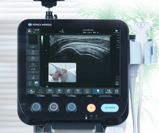
announced today several new solutions in digital radiography (DR) and ultrasound that will be introduced at the 2023 Radiology Society of North America (RSNA) Scientific Assembly and Annual Meeting from November 26-30 in Chicago, IL. milla1cf Sun, 11/26/2023 - 09:00 November 26, 2023 — Konica Minolta Healthcare Americas, Inc.,
milla1cf Thu, 11/23/2023 - 06:00 November 23, 2023 — Fujifilm Healthcare Americas Corporation, a leading provider of diagnostic and enterprise imaging solutions, is unveiling several new medical systems at the 2023 Radiological Society of North America ( RSNA ) annual meeting, booth #1929, held November 26 – 30 at McCormick Place in Chicago.
An AI algorithm was presented that could make dynamic digital radiography (DDR) more efficient by automatically measuring kinematics involved in certain shoulder injuries. performed well on image-independent American College of Radiology Diagnostic In-Training Exam (ACR DXIT) practice questions. Click here to watch the interview.
Chest radiography is a common diagnostic tool, but significant training and experience is required to interpret exams correctly,” said lead researcher Louis L. fellow in the Department of Radiology at Herlev and Gentofte Hospital in Copenhagen, Denmark. Plesner, M.D., resident radiologist and Ph.D.
Optimizing the use of equipment in the hospital can alleviate the burden on staff and patients. Confidence in non-radiological environments Another study to be presented at the Congress is “Dual-energy acquisition of portable chest X-rays: added diagnostic value in a non-radiological reviewing environment”.
Optimizing the use of equipment in the hospital can alleviate the burden on staff and patients. Confidence in non-radiological environments Another study to be presented at the Congress is “Dual-energy acquisition of portable chest X-rays: added diagnostic value in a non-radiological reviewing environment”.
Current diagnostics utilize plain radiographs and manual measurements, comparing the injured distal tibiofibular joint to the non-injured side. When left undiagnosed and/or untreated, they may lead to syndesmotic instability and subsequently post-traumatic ankle osteoarthritis, which can be challenging to diagnose, especially when subtle.
With advance planning and an emphasis on learning, your facility can perform imaging studies with the same standard of care as a large children’s hospital. With advance planning and an emphasis on learning, your facility can perform imaging studies with the same standard of care as a large children’s hospital.
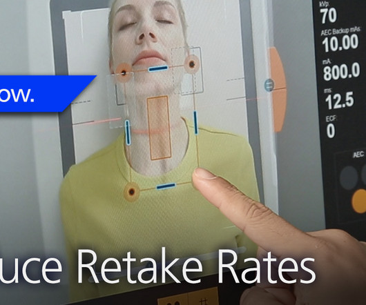
4) Precise patient positioning in radiology is essential to obtaining accurate diagnostic information to aid in effective patient care and for reducing a patient’s X-ray exposure due to retakes. 2 Unified Database for Rejected Image Analysis Across Multiple Vendors in Radiography.
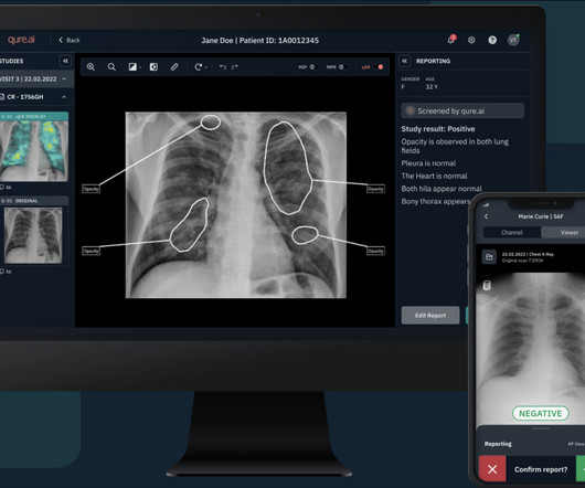
Qure’s chest X-ray based qXR-LN uses artificial intelligence to identify and localize lung nodules, marking another significant milestone for the organization, strengthening its standing as a pioneer in the realm of AI-powered advancements for plain film radiography and medical imaging. stands at the forefront of diagnostic advancement.
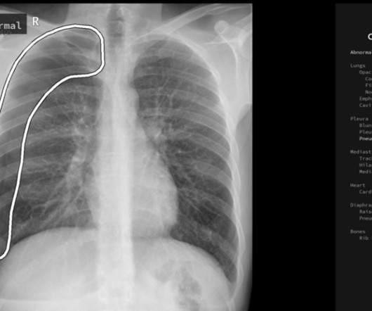
AI output Some hospitals have adopted digital portable X-ray machines to provide an instant image, the radiographer can see the chest X-ray immediately after exposure and decide whether the image quality is optimal.
Marion Anderson, CRA, Diagnostic Imaging Manager at Karmanos Cancer Center in Detroit, Michigan reports that her facility is down two-thirds of staff for CT and mammography exams. University Hospitals of Cleveland in Ohio undertook a thorough review of their compensation model to make sure they are competitive. “We
The Birth of X-ray Technology: At the end of the 19th century, Wilhelm Conrad Roentgen’s discovery of X-rays opened up new possibilities in medical diagnostics. As radiology departments proliferated in hospitals, X-rays became indispensable for diagnosing a wide range of conditions.
S hamie Kumar describes her perspective on how radiography has evolved over time, the impact radiographers can have in detecting abnormal X-rays and reflects how she views fast approaching AI in advancing current skills. The Red Dot System Often one of the first courses a newly qualified radiographer attends is the red dot course.
This transformative approach not only facilitates the interpretation of medical images but also transcends traditional boundaries, offering a comprehensive and insightful understanding of diagnostics. Traditional film-based radiography gives way to a digital ecosystem, enhancing the quality, accessibility, and shareability of medical images.
teleradiology India In the intricate tapestry of modern healthcare, teleradiology stands as a dynamic force behind the screens, reshaping the landscape of diagnostic imaging and patient care. Patients in remote areas gain access to specialized diagnostic services, contributing to equitable healthcare distribution.
Diagnostic Imaging Systems, Inc. Read More Understanding Sugar Levels in Horses’ Hay By: Clair Thunes, PhD Learn how to choose the right type of forage for horses with metabolic problems.
Powered by Eclipse, ImageView utilizes AI and Carestream’s proprietary algorithms to provide our most advanced image processing capabilities for superb image quality and diagnostic confidence. Physicians have better diagnostic confidence when they can view radiographs in the manner most suitable to their preferences.
It’s difficult, however, for many patients in a nursing home or other care environment to receive the appropriate diagnostic screenings in an efficient and comfortable manner. On top of the high costs associated with imaging services at a hospital , organizing each trip takes time and effort, then a staff member must accompany the patient.
Radiography has no role in orbital injuries due to its lower sensitivity for soft tissues [4]. 2015.01.004 Sai Kilaru is a medical student at Central Michigan University College of Medicine and plans to pursue a residency in diagnostic radiology. Lens subluxation can be diagnosed by ultrasound which shows deviation of the lens (Fig.
Coronavirus imaging from hospitals all over the world was collated to provide real-time COVID reporting best practice as the world started to understand the virus more. In addition, top thoracic specialist radiologists from Europe who had already experienced COVID radiology were called to report cases for NHS hospitals.
As a previous imaging director in the NHS for many years he developed a range of new services, most recently as lead radiologist for Royal Brompton HospitalDiagnostic Imaging Centre, opened in 2022. This facility incorporates one of the only combined interventional bronchoscopy and radiology facilities in the country.
No início desta primavera, John Crowther, gerente de marketing digital da Carestream, teve a oportunidade de viajar até o Hospital Shriners Children’s, localizado em St. Metas alinhadas As soluções da Carestream ajudam a proporcionar uma experiência aprimorada para os pacientes e a equipe do Shriner’s Children’s Hospital em St.
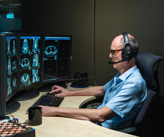
With mobile imaging, certified technologists and radiologists go directly to a medical facility, residence or placeof business with all of the necessary equipment needed to conduct a variety of diagnostic screenings. They would besent to an in-house imaging department, a third-party imaging facility or a hospital.
Is the diagnostic validity of conventional radiography for Lisfranc injury acceptable? She is now pursuing a career in Diagnostic Radiology with interests in Breast imaging. Treasure Island (FL): StatPearls Publishing; August 29, 2022. PMID: 28846306. Bookshelf ID: NBK448147. Chen C, Jiang J, Wang C, Zou J, Shi Z, Yang Y.
We organize all of the trending information in your field so you don't have to. Join 5,000 users and stay up to date on the latest articles your peers are reading.
You know about us, now we want to get to know you!
Let's personalize your content
Let's get even more personalized
We recognize your account from another site in our network, please click 'Send Email' below to continue with verifying your account and setting a password.


Let's personalize your content