This site uses cookies to improve your experience. To help us insure we adhere to various privacy regulations, please select your country/region of residence. If you do not select a country, we will assume you are from the United States. Select your Cookie Settings or view our Privacy Policy and Terms of Use.
Cookie Settings
Cookies and similar technologies are used on this website for proper function of the website, for tracking performance analytics and for marketing purposes. We and some of our third-party providers may use cookie data for various purposes. Please review the cookie settings below and choose your preference.
Used for the proper function of the website
Used for monitoring website traffic and interactions
Cookie Settings
Cookies and similar technologies are used on this website for proper function of the website, for tracking performance analytics and for marketing purposes. We and some of our third-party providers may use cookie data for various purposes. Please review the cookie settings below and choose your preference.
Strictly Necessary: Used for the proper function of the website
Performance/Analytics: Used for monitoring website traffic and interactions
The need for imagingmodalities to support an earlier, more accurate diagnosis continues to drive AI applications in the medical imaging market. In fact, the size of the market for AI in medical imaging is experiencing phenomenal growth, expected to increase from $1.12 billion in 2022 to $27.52
"Together with our radiographers, I learned to scan cardiac patients and learned special anatomy from pediatric cardiologists and pediatric cardiac surgeons." Since Gutberlet was starting out, training for cardiac imaging has improved. years of follow-up compared with CT imaging (2.1%
Substituting less energy-consuming ultrasound for x-ray or CT reduced energy use by as much as 8% during diagnostic radiology processes and 31.2% for total CFCs, while demonstrating an increase in diagnostic activity of 11.8%, Masperi said. Stage two consisted of a retrospective review of exams performed and their timing.
In the third blog of her series on AI and the radiographer, Shamie Kumar explores the impact on the radiographer when AI is integrated within an imagingmodality. The question to explore in this blog is when AI is integrated within an imagingmodality itself and how that may impact a radiographer.
The department of radiology teaches a diagnostic technologies in healthcare course, principally around imaging technologies, for which 3,000 people have signed up, Rubin said. "It
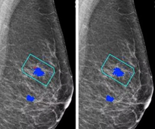
AI software based on deep-learning algorithms is showing promise, however, for helping to improve specificity in screening mammography and other breast imagingmodalities. Although mammogram is the most widely used screening modality, a known problem is that 9.5% Just over 40.5 million mammograms were performed in the U.S.
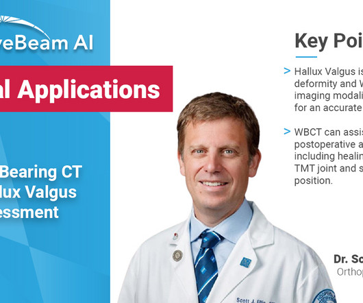
Key Points: The most important advantage of weight bearing CT (WBCT), which utilizes cone beam CT (CBCT) technology (a three-dimensional (3D) imagingmodality) is immediate access to 3D images, resulting in faster and better diagnostic capabilities.
The enduring shortage is affecting staffing for radiographers and radiologists; and all imagingmodalities. Marion Anderson, CRA, DiagnosticImaging Manager at Karmanos Cancer Center in Detroit, Michigan reports that her facility is down two-thirds of staff for CT and mammography exams. Bureau of Labor Statistics.
This blog delves into the captivating realm of radiology and the intricate art of crafting diagnosticimages with X-ray precision. The Canvas of Medicine: Radiographic Film and Digital Sensors: The canvas on which these diagnosticimages are painted has evolved.
Kim Mason Kim Mason, an Audit and Research Radiographer for Mid Yorkshire Teaching Hospitals Trust, talks about their role as well as the value of radiographer engagement in research activities and how to get involved. Hi, I’m Kim and I am an alternative-styled, funky-haired, septum-pierced, disabled Audit and Research Radiographer.
Key Points: Weight bearing CT (WBCT) can detect signs of osteoarthritis (OA), such as osteophytes, subchondral cysts, and joint space narrowing better than radiographs. Emerging evidence suggests WBCT reveals meniscal extrusion not detected by MRI and WBCT arthrography visualizes meniscal tears.
Reading Time: 4 minutes read Key takeaways from the 2023 RSNA Conference The topic of artificial intelligence technology trends in medical imaging was once again infused throughout sessions and on display in the exhibitor hall at the 2023 Radiological Society of North America (RSNA) scientific assembly and annual meeting.
Diagnosis While radiographs are typically sufficient to make the diagnosis, WBCT scans may be useful to plan surgical treatment. Accurately assess sesamoid position as plain radiographs cannot determine whether the sesamoids have been reduced within their grooves 5. Accurately assess healing in the 1st TMT joint 4. 10.30795/jfootankle.2022.v16.1674.
With advance planning and an emphasis on learning, your facility can perform imaging studies with the same standard of care as a large children’s hospital. Typically, one of two things may happen when an imaging technologist or sonographer who infrequently performs pediatric diagnosticimaging studies images a child.
after seeing the image. (2) Photoprint from radiograph by W.K. 3) In the early twentieth century, it was a common goal for investigators to try to find a way to separate the superimposed shadows that were recorded when a complex structure was shown on a radiograph. (3) 15) Radiologists needed a common means for sharing images.
rebound and guarding) Diagnostics Laboratory Tests Commonly ordered lab tests (i.e. There is no history or physical exam feature that rules out the disease Lactate elevation is a late finding in SBO. A normal lactate does not rule out the diagnosis Plain X-rays perform poorly in making or ruling out the diagnosis.
There is great value in comparing both ankles to reduce the chance of diagnostic error related to the large anatomical variability of the distal tibiofibular area. Overall, I see great value for WBCT in detecting “subtle” ligamentous injuries, which are those more likely to go unseen on traditional imaging.
For dense-breasted patients requiring supplemental imaging, MRI remains a valuable option that is not limited by breast density and is shown to be more sensitive than mammography at finding breast cancer. vi Investigations continue of this newer imagingmodality, which has the potential to positively benefit patients with dense breasts.
Key Points: Imagingmodalities such as plain radiographs (X-Ray), computed tomography (CT), and magnetic resonance imaging (MRI), dont have the diagnostic accuracy needed to detect syndesmotic widening or subtle instability. and threshold diagnostic values of relative syndesmotic widening as low as 0.43
In the world of orthopedic diagnostics, imaging plays a crucial role in identifying deformities and planning surgical interventions. Conventional radiographs and MRIs have been the standard, but they come with limitations when it comes to understanding the complex, three-dimensional structure of the human body. Why Choose WBCT?
What if X-ray imaging, the most prevalent and accessible imagingmodality in the world, could provide the information needed for diagnosis? The study could lead to radiographs providing an early window into disease manifestations that are currently undetected, thus leading to an earlier diagnosis of IPF.
3] To identify a causative vascular lesion, which may or may not be amenable or contraindicatory to thrombolysis Non-Contrast Head CT NCCT is usually the first imagingmodality obtained in the acute evaluation for stroke. Cerebral CT angiography and CT perfusion in acute stroke detection: a systematic review of diagnostic value.
Newborns' livers can be affected by a variety of congenital and acquired diseases, and imaging plays an important role in the workup and management of these, according to a study published November 7 in RadioGraphics. The team outlined the pros and cons of various modalities for neonatal liver imaging: X-ray. Ultrasound.
We organize all of the trending information in your field so you don't have to. Join 5,000 users and stay up to date on the latest articles your peers are reading.
You know about us, now we want to get to know you!
Let's personalize your content
Let's get even more personalized
We recognize your account from another site in our network, please click 'Send Email' below to continue with verifying your account and setting a password.


Let's personalize your content