This site uses cookies to improve your experience. To help us insure we adhere to various privacy regulations, please select your country/region of residence. If you do not select a country, we will assume you are from the United States. Select your Cookie Settings or view our Privacy Policy and Terms of Use.
Cookie Settings
Cookies and similar technologies are used on this website for proper function of the website, for tracking performance analytics and for marketing purposes. We and some of our third-party providers may use cookie data for various purposes. Please review the cookie settings below and choose your preference.
Used for the proper function of the website
Used for monitoring website traffic and interactions
Cookie Settings
Cookies and similar technologies are used on this website for proper function of the website, for tracking performance analytics and for marketing purposes. We and some of our third-party providers may use cookie data for various purposes. Please review the cookie settings below and choose your preference.
Strictly Necessary: Used for the proper function of the website
Performance/Analytics: Used for monitoring website traffic and interactions
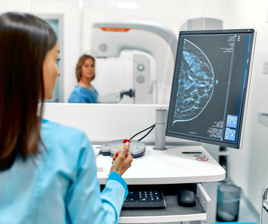
Mammograms are a crucial diagnostic tool that helps doctors detect early signs of breast cancer and other breast-related issues. The truth is mammograms are generally safe when used properly, and the amount of radiation you’re exposed to is minimal. Radiation exposure is controlled and minimized to ensure patient safety.
million mammograms were performed in the U.S. Although mammogram is the most widely used screening modality, a known problem is that 9.5% As is common in Europe, NHS currently uses a two-radiologist reader assessment of breast mammograms, three when there is disagreement. Just over 40.5
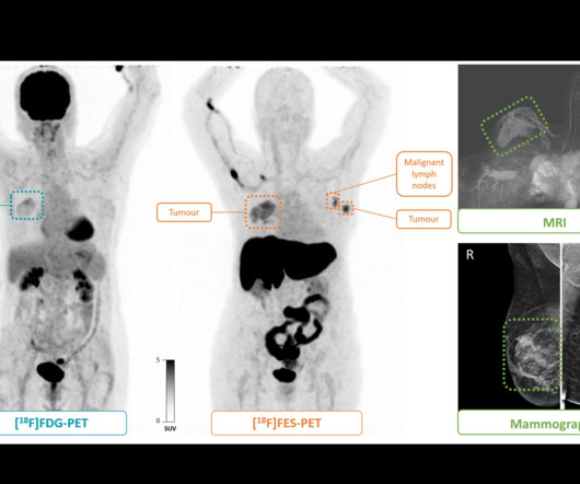
B) F-18 FDG-PET maximum intensity projection, F-18 FES-PET maximum intensity projection, breast MRI scan, and mammogram in an 81-year-old female participant who presented with a tumor in the right breast. All lesions had also been identified at mammography and MRI. Image and caption courtesy of the RSNA.
Breast density can often obscure lesions on conventional x-ray mammography, and so other screening modalities such as MRI or ultrasound are often recommended for follow-up. CEM is faster and less costly than MRI and can often be used as a follow-up to an abnormal screening mammogram when it is clinically appropriate.
Mortazavi and colleagues evaluated data from 23 women who presented with axillary adenopathy on mammography, breast ultrasound, or breast MRI after being vaccinated for COVID-19 between December 2020 and February 2021. Ultrasound from diagnostic work-up performed seven days later showed no change in lymph node size.
Komen has urged quick passage in Arizona of legislation that would eliminate patient out-of-pocket costs for diagnostic and supplemental breast imaging. House Bill 2411 was introduced in the state by Representative David Cook (R-Globe) and includes eliminating costs for patients for MRI, ultrasound, and diagnosticmammograms.
The reduction for the bilateral mammogram 77066 was 1.36%, reflecting an increase in RVU valuation that somewhat offsets the conversion factor cut. Effect on professional component reimbursement The single-view chest x-ray 71045 professional fee was cut 5.55%.
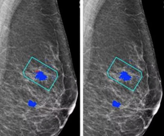
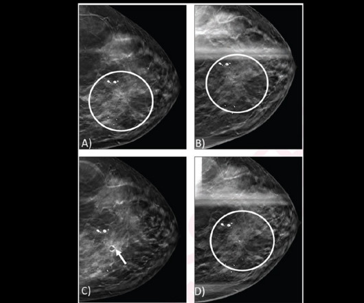
A) Craniocaudal view from screening DBT mammogram shows architectural distortion (circle) in the upper inner position, which was not detected by digital mammography (not shown). (B) B) Spot craniocaudal view from subsequent diagnostic DBT mammogram shows persistence of architectural distortion (circle).
CNNs have been used in AI research to classify breast cancer disease status and predict therapeutic responses based on diagnostic images of the primary breast tumor. The researchers highlighted that CNNs require little preprocessing compared with other image classification algorithms. DEED Attribution-NonCommercial-NoDerivs 4.0 International.
The American Cancer Society recommends starting annual mammogram screenings at age 40. Mammogram Screening Mammogram: Screening mammograms take 2 or more images of each breast. DiagnosticMammogram: DiagnosticMammograms are ordered for a variety of reasons and may assess one or both breasts.
That changed in 2023, when Angie underwent a mammogram and breast ultrasound at a Midstate Radiology Associates location offering CARE. Based on Angie’s risk score, she qualified for a breast MRI, a diagnostic tool capable of detecting cancers that mammograms and ultrasounds might miss.
A radiologist’s perception when viewing a complex MR image may be akin to a Major League Baseball (MLB) batter reading the stitches on a fastball, according to researchers exploring exactly how diagnostic interpretations are made.
Why Breast Density Matters in Cancer Screening Dense breast tissue affects screening in two key ways: Reduced Visibility : Dense tissue appears white on mammograms, as do tumors, making it harder to detect abnormalities. Inter-radiologist Variation : Assessments can vary up to 33% 1 when different radiologists interpret the same mammograms.
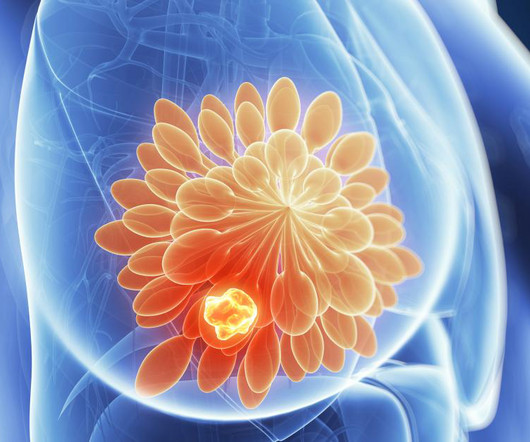
The earlier breast cancer is detected through diagnostic imaging, the better chance there is for successful treatment with surgery, radiation therapy, or chemotherapy. Consistent mammography and diagnostic imaging appointments can help stop this number from continuing to rise. this year alone. this year alone.
Capitol Imaging Services has served tens of thousands of women with our women’s health examinations in screening mammography, diagnostic mammography, ultrasound, bone density studies, breast MRI and breast biopsy. Our independent facilities streamline our processes because screening and diagnostic testing is all we do.
The increased specificity of follow-up CEM examinations compared with baseline CEM examinations suggests that the diagnostic performance of CEM may be improved even further in the future,” Jochelson and colleagues wrote. From the CEM exams, the team defined "low-energy" images as the equivalent of a 2D full-field digital mammogram.
Another waited three months for mammogram findings. Theyre part of a larger trend, driven by a persistent imbalance between the number of radiologists available and the ever-growing demand for diagnostic imaging. In Michigan, a patient recently reported waiting over 80 days for imaging results. These delays arent isolated.
“In recent years, AI has been studied for the purpose of diagnosing breast cancer earlier by automatically detecting breast cancers in mammograms and measuring the risk of future breast cancer.” Diagnostic AI models are trained to detect suspicious lesions on mammograms and are well suited to estimate short-term breast cancer risk.
For dense-breasted patients requiring supplemental imaging, MRI remains a valuable option that is not limited by breast density and is shown to be more sensitive than mammography at finding breast cancer. Diagnostic Imaging Utilization in the Emergency Department: Recent Trends in Volume and Radiology Work Relative Value Units.
With three outpatient imaging locations throughout Boise, Meridian, and Eagle, Intermountain Medical Imaging offers diagnostic imaging procedures in a calm and comforting environment, including MRI, CT, Ultrasound, X-ray, Interventional Radiology, and Mammography. “St. This new partnership between St.
Since almost half of the screening population has dense breasts, many of these patients require additional breast imaging, often with MRI , after mammography. Low-dose positron emission mammography (PEM) is a novel molecular imaging technique that provides improved diagnostic performance at a radiation dose comparable to that of mammography.
Techniques such as mammography, low-dose computed tomography (LDCT), and magnetic resonance imaging (MRI) are instrumental in identifying cancers like breast, lung, and prostate in their nascent stages. At Vesta Radiology , we are committed to leveraging the advantages of teleradiology to provide exceptional diagnostic services.
In addition to mammograms and ultrasounds, women should know about the various other breast screenings that are performed. Diagnostic Mammography Diagnosticmammograms may be performed when a woman (or man) has presented with some type of symptom that requires further examination.
Try as we might, good health practices tend to be neglected, especially regarding preventive health screenings like mammograms. Research shows that one in four women who should be getting regular mammograms don’t. Women with an average risk for breast cancer should begin mammogram screening every year starting at 40.
Women with dense breasts are BOTH more likely to develop breast cancer and more likely to have that cancer missed on a mammogram [5] Fig. 1 – Cancer on a mammogram of a fatty vs a dense breast What is Dense Breast Tissue? Breast density is determined through a mammogram and described as one of four categories (Fig.
MRI-Scan-Teleradiology Introduction: Mammography is not just a diagnostic tool; it’s a mastery of precision and compassion in breast imaging. This blog is a journey through the world of mammography, shedding light on the importance of precision and compassion in breast health.
A doctor will often use a diagnostic imaging test to check patients for signs of cancer. There are several types of imaging tests that physicians use to detect cancer in patients: X-Ray, Computed Tomography (CT), Magnetic Resonance Imaging (MRI), Ultrasound (US), Nuclear Medicine, and Positron Emission Tomography (PET).
Medical Imaging of Fredericksburg’s services include X-Ray, CT, PET/CT, MRI, 3D mammograms, Ultrasound and a variety of other health scans. Patients have given an over 95% satisfaction score – a reflection of the level of commitment the physicians and staff have to the community they serve.
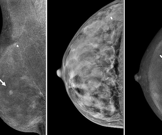
A) Mammogram MLO view. Mammogram CC view. A mammogram demonstrated focal asymmetries involving most of the anterior and mid right breast with diffuse skin thickening, trabecular coarsening, increased overall density, and enlarged right axillary lymph nodes. What is the diagnosis? Xray of the Week Figure 1. Surg Clin North Am.
All of the annual scheduled services such as mammograms can now be scheduled, as well as imaging prescribed by physicians for the care of their patients. Patients may also schedule mammograms directly at our facilities in these same locations. What this means for our community and region is important.
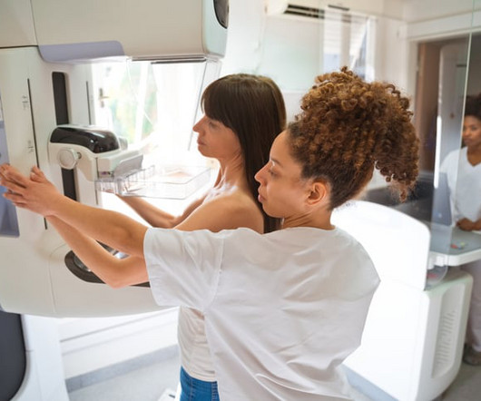
When mammography is carried out on an individual without any symptoms, it is called screening mammography whereas if carried out on an individual with symptoms, it is called diagnostic mammography in which additional views have to be carried out. If the breast is dense on the mammogram, an ultrasound must also be carried out.
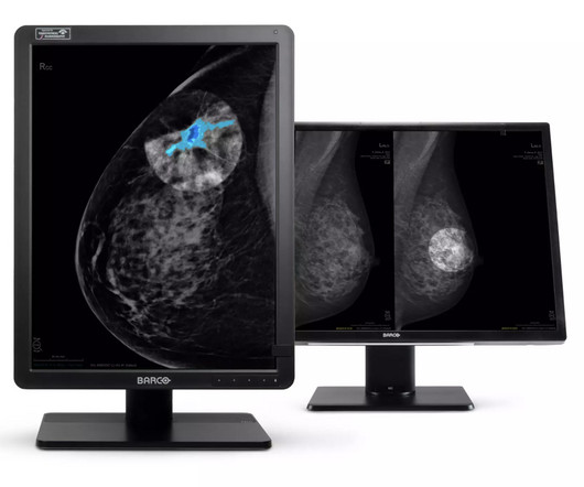
“The existing Intuitive Workflow Tools will benefit from this add- on AI software tool which addresses a significant unmet need in medical imaging, enabling rapid and accurate lesion segmentation, measurement, and visualization” says Dirk Feyants , Executive Vice President of Diagnostic Imaging at Barco.
Whether you’re looking for a second set of eyes for complex cases or want to ensure the highest level of diagnostic accuracy, our team of board-certified radiologists—with subspecialties in areas such as neuroradiology, musculoskeletal imaging, and oncology—are ready to assist. Why Choose a Teleradiology Partner for Second Opinions?
Therefore, it makes sense to have Mammo Techs performing breast ultrasounds, as well as mammograms. It was agreed upon that our diagnostic technologists would become breast ultrasound certified. what to image for documentation and when to refer to CESM, MBI, or MRI).
The use of comprehensive imaging using co-registered functional and morphological information can reduce reading time to seven – ten minutes versus thirty to sixty minutes for a standard breast MRI [4] , [5]. Diagnostic innovation is on a trajectory that we cannot ignore. What Is a Breast MRI? Breast Cancer Screening.
a screening mammogram). With locations from Texas to Florida, getting the highest-quality MRI, CT, ultrasound or other type of exam is convenient and yet may cost much less. Ask about your options for free preventative care. Check with your HR department or your doctor about what care is available to you.
An MCQ that asks the learner to recognize benign dermal calcifications on a mammogram does not test the learner’s problem-solving ability or ability to communicate the findings to a patient. For example, they are not the best way to test a radiologist’s ability to communicate effectively.
What’s more, the USPSTF concluded that there was insufficient evidence to recommend supplemental screening with MRI or ultrasound in women, regardless of breast density. Furthermore, high-risk women who desire supplemental screening -- but cannot undergo MRI -- should consider contrast-enhanced mammography, according to the ACR.
Studies have revealed inequalities in access to 3D mammography facilities and delays in obtaining diagnostic evaluations, which undermine the benefits of early cancer detection. There’s also a need for more research on supplemental screening modalities, such as ultrasound or MRI, for women with dense breasts.
To continue improving outcomes, mammograms and regular screenings remain our best tools to catch breast cancer before it becomes life-threatening. Regular mammograms, breast self-exams, and clinical breast exams can help identify abnormalities before the disease progresses.
In the 1960s and 1970s, scientific research was published about the diffusion, relaxation and chemical exchange of water intracellularly, eventually leading to Magnetic resonance imaging (MRI). (6) Their work gave rise to the modern MRI scanners we use today. He named this the focusing NMR concept (FONAR). (8) doi: 10.1126/science.171.3976.1151
This included changes in the ages at which women were recommended to start getting annual mammograms or all adults were recommended to get colonoscopies or other tests for colorectal cancer, as well as the introduction in 2013 of CT scan-based screening for lung cancer in certain current or past smokers.
We organize all of the trending information in your field so you don't have to. Join 5,000 users and stay up to date on the latest articles your peers are reading.
You know about us, now we want to get to know you!
Let's personalize your content
Let's get even more personalized
We recognize your account from another site in our network, please click 'Send Email' below to continue with verifying your account and setting a password.


Let's personalize your content