This site uses cookies to improve your experience. To help us insure we adhere to various privacy regulations, please select your country/region of residence. If you do not select a country, we will assume you are from the United States. Select your Cookie Settings or view our Privacy Policy and Terms of Use.
Cookie Settings
Cookies and similar technologies are used on this website for proper function of the website, for tracking performance analytics and for marketing purposes. We and some of our third-party providers may use cookie data for various purposes. Please review the cookie settings below and choose your preference.
Used for the proper function of the website
Used for monitoring website traffic and interactions
Cookie Settings
Cookies and similar technologies are used on this website for proper function of the website, for tracking performance analytics and for marketing purposes. We and some of our third-party providers may use cookie data for various purposes. Please review the cookie settings below and choose your preference.
Strictly Necessary: Used for the proper function of the website
Performance/Analytics: Used for monitoring website traffic and interactions
Women in racial and ethnic minority backgrounds are less likely to be provided same-day diagnostic breast imaging services, despite such services being available, according to research published February 18 in Radiology. Additional imaging and possibly image-guided biopsy are recommended for women who have an abnormal screening mammogram.
million mammograms were performed in the U.S. Although mammogram is the most widely used screening modality, a known problem is that 9.5% Starting with mammography images, the work is important for finding ways to alleviate the region's radiologist workforce shortage and staffing pressures, according to Imperial College London.
A radiologist’s perception when viewing a complex MR image may be akin to a Major League Baseball (MLB) batter reading the stitches on a fastball, according to researchers exploring exactly how diagnostic interpretations are made. They found that some observers performed better than expected for their rank and years of experience.
Maybe you recently decided to try the best 3D mammogram experience in El Paso, have recently moved, changed doctors, or acquired new insurance, and are now going to our imaging center for your annual mammogram. In this case, the radiologist may recommend a diagnosticmammogram. How Can I Obtain My Prior Images?
While the pandemic affected medical operations across the country, the experts said that radiologists developed and honed their sense of resiliency as imaging was placed on the front lines. While screening mammography services were halted, Patel and colleagues continued with diagnostic imaging services, especially for symptomatic patients.
To do so, three radiologists annotated a dataset of mammograms using histology-based ground truth. To do so, three radiologists annotated a dataset of mammograms using histology-based ground truth. These networks were also validated and tested using an annotated dataset of 1,000 patients and 1,986 mammograms.
milla1cf Mon, 05/22/2023 - 13:25 May 22, 2023 — Incorrect advice by an AI-based decision support system could seriously impair the performance of radiologists at every level of expertise when reading mammograms, according to a new study published in Radiology , a journal of the Radiological Society of North America ( RSNA ).
A deep-learning algorithm can rule out the presence of breast cancer on screening mammograms, improving specificity and yielding significant workflow and downstream savings, according to research published April 10 in Radiology. dataset 1: 143,593 mammograms interpreted by 11 breast radiologists from 2008 to 2017 U.S.
We're excited to introduce advanced workstation features for our flagship solution, ProFound Detection, aimed at further improving and facilitating radiologists' interpretation of mammograms within their workstation,” said Dana Brown , President and CEO of iCAD.
Healthcare disparities continue to plague medical imaging, but there are concrete measures radiologists can take to mitigate them, according to a paper published on October 12 in RadioGraphics. B) A craniocaudal magnified mammogram more clearly shows the irregular mass and pleomorphic calcifications in the medial breast. (C)
Breast density assessment is important not only for the patients health, but it also has reimbursement implications for radiologists. CEM is faster and less costly than MRI and can often be used as a follow-up to an abnormal screening mammogram when it is clinically appropriate.
As more people get vaccinated for COVID-19, radiologists must be familiar with how the vaccine may affect imaging results, wrote Shabnam Mortazavi, MD, of the University of California, Los Angeles. insert table here) (Above) 55-year-old woman who underwent screening mammogram and ultrasound seven days after first COVID-19 vaccination dose.
The original ScreenTrustCAD trial (2021–2022), published in The Lancet Digital Health , was the first prospective interventional study to demonstrate that AI could match or even surpass traditional two-radiologist systems. These findings demonstrate AI’s ability to enhance diagnostic accuracy and significantly improve screening efficiency.
christine.book Tue, 05/21/2024 - 10:36 May 21, 2024 — According to a newly-published study of nearly 5,000 screening mammograms interpreted by an FDA-approved AI algorithm, patient characteristics such as race and age influenced false positive results. Duke Health , breast radiologist and assistant professor at Duke University in Durham, NC.
The Benefits of Regular Mammograms for Women Mammography plays a crucial role in breast cancer prevention by contributing to early detection, which is one of the most effective ways to reduce mortality and improve treatment outcomes. Schedule your mammogram appointment today at (915) 225-2480 ! They are considered precancerous changes.
Methods for training artificial intelligence (AI) diagnostic algorithms can help prevent these models from producing medical errors caused by adversarial attacks, according to research presented at RSNA 2023 in Chicago. Adversarial data produced by projected gradient descent were used to insert noises into mammogram images.
One question our technologists are asked frequently is, “What’s the difference between a diagnosticmammogram and a screening mammogram?” Screening Mammogram A Screening Mammogram is a routine x-ray exam for women who have not experienced any symptoms or abnormalities with their breasts.
Lunit's AI assigns each breast a score from 0 to 100, with higher values indicating a greater likelihood of cancer being present on the current mammogram. The double reading of mammograms, though the standard of care in Denmark and other European regions, is resource-intensive and demands significant radiologist time.
Saige-Dx optimizes breast cancer screening to help radiologists detect even subtle lesions, according to the company. Saige-Dx is intended to be used on women 35 years old and older, and it is not intended to replace a physician's own review of a mammogram. It assists in a recall or don't recall decision.
Everything we do at DeepHealth is about empowering radiologists and healthcare professionals, not just in the detection and diagnosis of diseases but across the whole care continuum. Saige Breast has been trained on over 10,000 scans and has now been employed in over a million mammograms. What is DeepHealth doing this year at ECR?
Why Breast Density Matters in Cancer Screening Dense breast tissue affects screening in two key ways: Reduced Visibility : Dense tissue appears white on mammograms, as do tumors, making it harder to detect abnormalities. Inter-radiologist Variation : Assessments can vary up to 33% 1 when different radiologists interpret the same mammograms.
Artificial Intelligence (AI): Revolutionizing Radiology in 2025 AI continues to make waves in radiology, offering improved diagnostic accuracy and efficiency. In 2025, AI tools are more refined than ever, assisting radiologists with cancer detection, anomaly identification, and image interpretation.
With AI-mediated cyberattacks on the rise, radiologists can find it challenging to balance prompt patient access to diagnostic imaging while safeguarding sensitive healthcare data. With growing radiology practices and outpatient imaging facilities, we will see an increase in outsourced diagnostic imaging. Dhaval Shah.
CNNs have been used in AI research to classify breast cancer disease status and predict therapeutic responses based on diagnostic images of the primary breast tumor. Finally, two expert radiologists isolated the regions of interest of the primary tumor from other areas of breast tissue. DEED Attribution-NonCommercial-NoDerivs 4.0
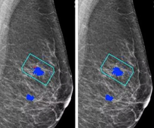
Topics include the following: Lunit's AI model combines new and existing AI algorithms to filter normal chest radiographs from the radiology workload An exploration of the accuracy and robustness of Lunit's Insight MMG compared with radiologist readers Lunit's AI model tracks mammographic parenchymal patterns as predictive markers for breast cancer (..)
Maybe you have recently moved, changed doctors, or have new insurance and now, are going to a different imaging center for your annual mammogram. Either way, the new imaging center will very often request that you provide the images from your previous mammograms. In this case, the radiologist may recommend a diagnosticmammogram.
As a teleradiology company, we specialize in bridging this gap by offering high-quality diagnostic imaging interpretation, ensuring rural healthcare providers can deliver top-tier care to their patients. Radiologists must inform patients if they have dense breast tissue, a factor that can obscure mammogram results and increase cancer risks.
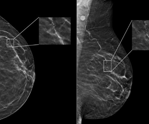
A new AI system that mimics the gaze of radiologists interpreting medical images, such as mammograms, can enhance the speed, precision, and sensitivity of medical diagnostics while facilitating early detection of breast cancer.
A new AI system that mimics the gaze of radiologists interpreting medical images, such as mammograms, can enhance the speed, precision, and sensitivity of medical diagnostics while facilitating early detection of breast cancer.
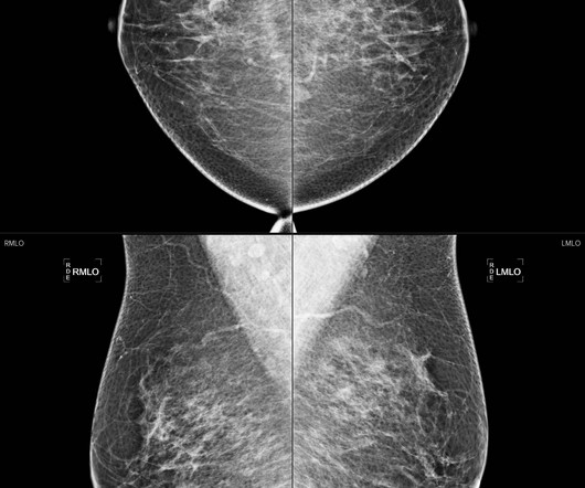
Screening Mammograms have proven to be essential for the early detection of breast cancer. What Is A Screening Mammogram? A mammogram is an examination that uses a special low dose X-Ray machine to evaluate breast tissue. The mammogram unit is designed specifically for breast imaging. Are Screening Mammograms Risky?
A combination of canceled elective screenings and procedures, staff and PPE shortages, office closures, and personal health concerns all contributed to a decline in the number of routine mammograms provided for at-risk women. Why do I need a Mammogram Every Year? The result is a rise in breast cancer diagnoses.
A swollen or enlarged lymph node identified during a mammogram can be a sign of breast cancer or lymphoma. If this timeline is not possible, patients should continue to attend all recommended appointments and be prepared to provide information about their recent vaccine before a mammogram. Why Are Swollen Lymph Nodes Concerning?
This technology mitigates the challenges posed by a shortage of on-site radiologists and enhances the quality of care in remote areas. A report from Healthcare IT News highlights how teleradiology enables radiologists to interpret scans remotely, increasing flexibility in work schedules and expanding access to specialized expertise.
Women who get that report saying they have dense breasts and that the mammogram doesn't work as well for them are kind of left in a little bit of a quandary," she told AuntMinnie.com. "We We really want people to speak to their primary care doctor, speak to their radiologist and make a joint decision about for their individual situation."
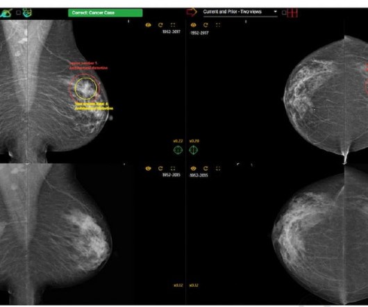
While researchers noted no significant impact on sensitivity rates, they found that access to a patient’s prior mammograms resulted in a nearly 15 percent increase in sensitivity for current mammography interpretation.
With the latest high-end medical display technology from LG, physicians can analyze radiological and mammogram images with complete confidence. LG Diagnostic Monitor (Model 32HQ713D 8MP Resolution) The LG Diagnostic Monitor. With the LG Digital X-Ray Detector, radiologists and physicians can examine clear x-ray images, fast.
With the onboarding of Johns Hopkins, Transpara's growing utilization in the northeast will now benefit radiologists and patients in over 50 sites throughout 5 states (NY, NJ, PA, MD, VA) and the District of Columbia. Transpara is designed to work concurrently with radiologists.
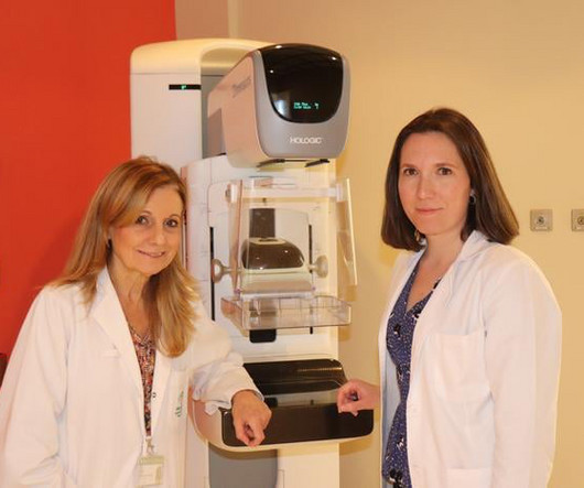
Despite the fact that this technique has become widespread in diagnostic units, it is still not as standardized in screening programs," says Marina Alvarez , head of the Radiodiagnostic Unit at the Reina Sofía Hospital, one of the authors of the study and a nationally prominent figure advocating for the early diagnosis of breast cancer.
mtaschetta-millane Mon, 07/29/2024 - 11:16 July 29, 2024 — Lunit, a leading provider of AI-powered solutions for cancer diagnostics and therapeutics, announced the implementation of its AI-powered breast cancer detection solution, Lunit INSIGHT MMG , in Qatar's national breast cancer screening program.
“In recent years, AI has been studied for the purpose of diagnosing breast cancer earlier by automatically detecting breast cancers in mammograms and measuring the risk of future breast cancer.” Diagnostic AI models are trained to detect suspicious lesions on mammograms and are well suited to estimate short-term breast cancer risk.
Utilizing Lunit INSIGHT MMG , an FDA -cleared and CE-marked AI solution for mammography analysis , this research backs AI's potential to replace one human reader under Europe's double reading guideline and underscores its capacity to alleviate the strain on radiologists. 4.44% decrease) and when operating independently (RR 1.55, 47.1%
For example, in Sweden, AI shortened the time-to-treatment for patients with incidental pulmonary embolism (iPE) by 97%, flagging suspected positive CT scans for radiologists to review as a priority. Current problems in diagnostic radiology vol. 7 AI can help alleviate some of this pressure. 51,4 (2022): 556-561.
Health equity training can help future radiologists better understand diverse patient needs, according to results published February 8 in Clinical Imaging. Imaging leaders have emphasized the need to recognize barriers patients and radiologists face to provide equitable access and quality care.
s national network of premier providers of diagnostic imaging services located in Western New York. This technology helps to augment radiologists by providing a concurrent read of screening and diagnosticmammograms, often enhancing early detection of breast cancer. “We
We organize all of the trending information in your field so you don't have to. Join 5,000 users and stay up to date on the latest articles your peers are reading.
You know about us, now we want to get to know you!
Let's personalize your content
Let's get even more personalized
We recognize your account from another site in our network, please click 'Send Email' below to continue with verifying your account and setting a password.


Let's personalize your content