This site uses cookies to improve your experience. To help us insure we adhere to various privacy regulations, please select your country/region of residence. If you do not select a country, we will assume you are from the United States. Select your Cookie Settings or view our Privacy Policy and Terms of Use.
Cookie Settings
Cookies and similar technologies are used on this website for proper function of the website, for tracking performance analytics and for marketing purposes. We and some of our third-party providers may use cookie data for various purposes. Please review the cookie settings below and choose your preference.
Used for the proper function of the website
Used for monitoring website traffic and interactions
Cookie Settings
Cookies and similar technologies are used on this website for proper function of the website, for tracking performance analytics and for marketing purposes. We and some of our third-party providers may use cookie data for various purposes. Please review the cookie settings below and choose your preference.
Strictly Necessary: Used for the proper function of the website
Performance/Analytics: Used for monitoring website traffic and interactions
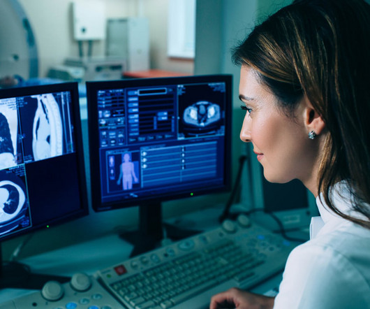
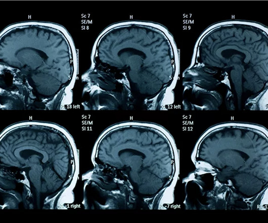
Deep learning-accelerated brain MRI improves the detection of acute ischemic lesions compared to conventional MRI because it offers better image quality and significantly decreased exam times, researchers have reported in a study published in Radiology. Arrows indicate the acute infarction.
Are low-field MRI units effective for neuroradiologic imaging? Use of low-field MRI for this indication would improve patientcare by expanding access to the modality, wrote a team led by medical student Lauren Kelsey. MRI for diagnosis and assessment of neurologic conditions is most commonly performed on 1.5-tesla
Middlebrooks' research interest consists of using ultrahigh-field, 7-tesla MRI to plot brain microstructure and develop surgical treatment of brain tumors, epilepsy, and neurodegenerative and movement disorders such as Parkinson's disease, essential tremor, and dystonia. Middlebrooks, MD, of the Mayo Clinic in Jacksonville, FL.
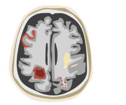
Example of how ARIA can present itself on a brain MRI scan. The FDA’s nod for icobrain aria comes at a time when much-needed new Alzheimer’s treatments are reshaping care options in the United States. Slow Adoption of Alzheimer’s Treatments Amid Safety Concerns Figure 1. icobrain aria was thoroughly evaluated in large reader studies.
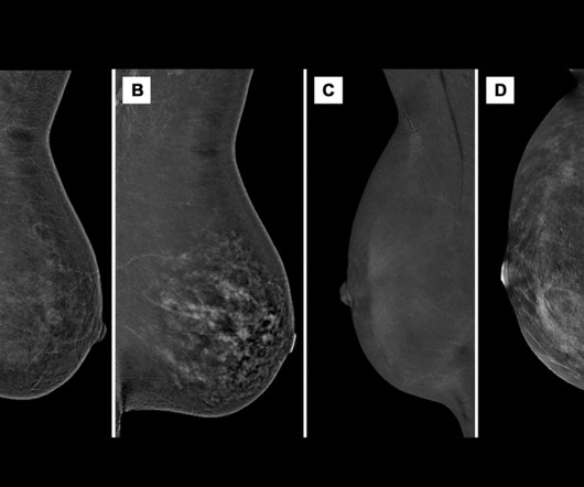
Our results highlight higher BPE in premenopausal compared with post-menopausal female patients, increased BPE during lactation and hormone replacement therapy, and decreased BPE during tamoxifen therapy, which increased at treatment cessation, Nissan and co-authors wrote. The full study can be accessed here.
were interpreted within the ordering provider's practice -- results that could have negative ramifications for patientcare, according to study coauthor Vijay Rao, MD, of Thomas Jefferson University in Philadelphia. for primary care physicians; 75.7% An HPI team found that 43.6% Ultrasound 52% Nuclear medicine 39.5%
Using AI software with brain MR imaging improves the diagnostic accuracy for the monitoring of amyloid-related imaging abnormalities (ARIA) in patients undergoing beta amyloid-directed antibody therapies for Alzheimer's disease, researchers have found. Among the patients, most (52.8%) were women and most (78.9%) were white.
His research interests include using structural and functional MRI -- particularly ultrahigh-field, 7-tesla MRI -- to map brain microstructure and develop neurosurgical treatment of brain tumors, epilepsy, and neurodegenerative and movement disorders such as Parkinson's disease, essential tremor, and dystonia. He served in the U.S.
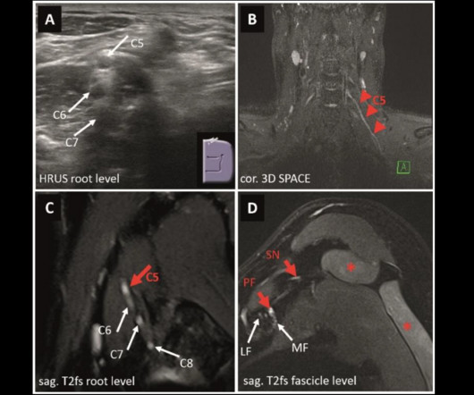
MRI and ultrasound have tradeoffs in diagnosing peripheral neuropathies of the upper extremity, a study published March 4 in Radiology found. However, the researchers noted that the diagnostic role of such imaging remains unclear due to limited clinical evidence. The full study can be accessed here.
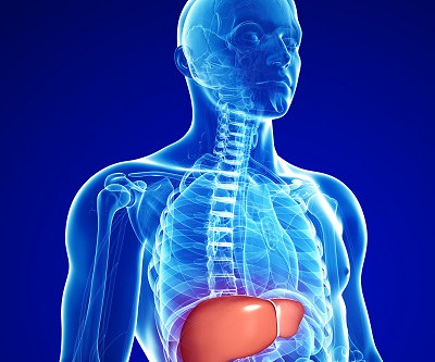
The MRI PDFF measure could improve patientcare by giving clinicians a more accurate picture of liver disease, a team led by Tianyi Xia, MD, of Zhongda Hospital School of Medicine, Southeast University, in Nanjing, China. There are a variety of ways to assess liver fat content, but MRI PDFF has shown high diagnostic precision.
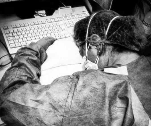
With 45% of radiologists reporting symptoms of burnout, Philips’ innovations across diagnostic imaging and enterprise informatics are focused on freeing up time for clinical staff through enhanced workflows and improved efficiency, according to information shared by the company during the conference. million liters of helium since 2018.
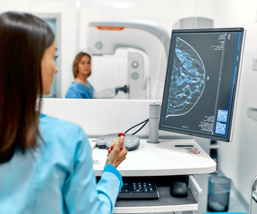
Mammograms are a crucial diagnostic tool that helps doctors detect early signs of breast cancer and other breast-related issues. However, many patients have concerns about radiation exposure and the potential risks involved. As the leading diagnostic imaging radiology center in El Paso, patientcare and safety are our top priorities.
A "CT first" strategy is an effective way to determine if patients with stable chest pain need revascularization with invasive coronary angiography (ICA), researchers have found.
Variability in the quality and interpretation of multiparametric MRI (mpMRI) prostate scans is a problem that confronts radiologists. Because mpMRI is widely used, radiology software developers have sought to improve and eliminate the drawbacks, especially to reduce the risk of errors for less skilled MRI readers and nonradiologists.
This non-invasive procedure is ideal for patients who experience chest pains or heart conditions, offering an alternative to invasive diagnostic techniques. Detect Abdominal and Pelvic Conditions For patients with abdominal pain, gastrointestinal issues, or similar abnormalities, CT scans can help identify these issues.
Reeder has been faculty at UW-Madison since 2005 and previously served as director of the clinical magnetic resonance imaging fellowship, chief of MRI, chief of sectional cardiovascular imaging, and senior vice chair of research. Reeder's term becomes effective in early 2024, according to UW.
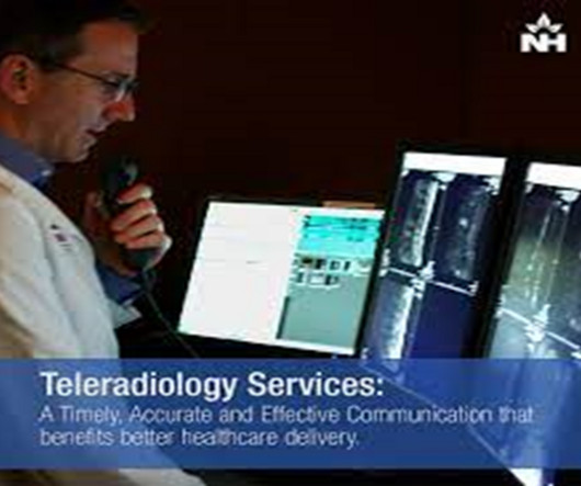
Teleradiology Introduction: Magnetic Resonance Imaging (MRI) stands as a radiant beacon in the realm of modern healthcare, continually illuminating new pathways to diagnostics and patientcare. In this exploration, we will shed light on how MRI is sparking a revolution, transforming diagnostics and patientcare.
The state of Washington is considering a bill authorizing diagnostic radiologic technologists (RTs), therapeutic RTs, and MRI technologists to perform intravenous contrast procedures under "general supervision," rather than direct supervision of a physician.
. | W3-SSNR10-1 | Room E353C In this scientific session, researchers will present study findings that suggest that a deep learning-based fast MRI reconstruction model improves the efficiency and quality of brain MRI exams in both spin-echo and gradient-echo sequences. on the rating scale compared with 3.4 for artifacts; and 0.55
Reeder has been faculty at UW-Madison since 2005 and previously served as director of the clinical magnetic resonance imaging fellowship, chief of MRI, chief of sectional cardiovascular imaging, and senior vice chair of research. Reeder's term becomes effective in early 2024, according to UW.
Introduction: In the realm of patientcare, MRI professionals play a crucial role in shaping diagnoses and improving healthcare outcomes. In this comprehensive exploration, we’ll delve into the essential functions of MRI professionals, highlighting their contributions to the healthcare ecosystem.
Managed by the American Medical Association (AMA), CPT codes are essential for documenting and billing medical, surgical, and diagnostic services. Source: AMA Press Release MRI-Guided High-Intensity Focused Ultrasound (MRgFUS) Non-invasive treatments for conditions such as intracranial disorders have received updated codes.
Medical School: They then complete four years of medical school, mastering the fundamentals of anatomy, pathology, pharmacology, and patientcare. This rigorous training covers all imaging modalities, from X-rays to advanced techniques like MRI and PET/CT scans.
AI is revolutionizing medical imaging across every aspect of diagnostic imaging as a triple threat in planning, scanning, and diagnosis. AI is not intended to replace the clinician or patientcare. It is the main tool for diagnostic imaging in oncology, neurology, and cardiology. Kelly Londy of GE HealthCare.
Theyre part of a larger trend, driven by a persistent imbalance between the number of radiologists available and the ever-growing demand for diagnostic imaging. Even with moderate increases in the number of new residents entering the field, demand for imaging especially advanced modalities like CT and MRI is expected to outpace supply.
As technological capabilities advance across all medical disciplines, so to do the imaging needs that often come with adequate diagnostic capabilities. Many operations managers find themselves wondering what equipment their facility needs to provide adequate services to their patient base.
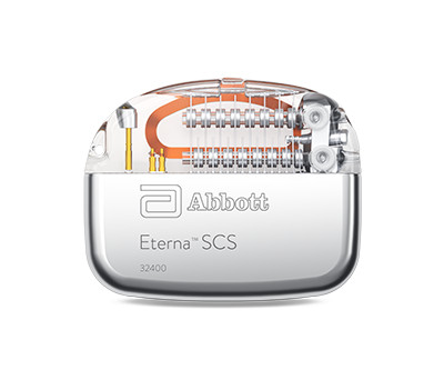
Food and Drug Administration (FDA) has approved expanded MRI labeling for its Eterna spinal cord stimulation (SCS) system to include new leads that are MR conditional, which means people with chronic pain can undergo MRI scans within the approved outlined conditions and have a wider selection of lead options for full-body scans.1,2
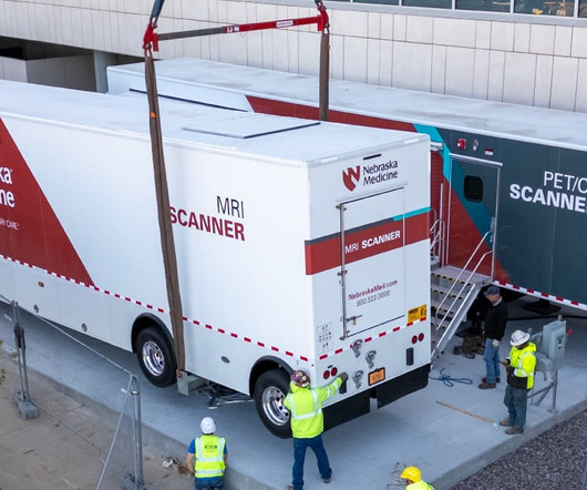
The MR 5300 with BlueSeal technology is designed to provide helium-free MR imaging capabilities in a mobile format, increasing access to care with advanced medical diagnostics for communities in both rural and urban areas. Both organizations strive to be the best and bring the best to the patients.”
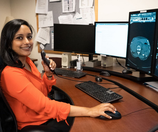
Techniques such as mammography, low-dose computed tomography (LDCT), and magnetic resonance imaging (MRI) are instrumental in identifying cancers like breast, lung, and prostate in their nascent stages. This approach addresses staffing challenges and enhances patientcare by providing timely, expert interpretations.
MRI Scanner clinical applications make it an invaluable asset in modern medical imaging GE Signa Artist Overview The GE Signa Artist 1.5T MRI Scanner is an advanced magnetic resonance imaging system designed to deliver high-quality imaging and exceptional patientcare. MRI Scanner first appeared on Amber Diagnostics.
Basics of MRI provide essential knowledge for understanding advanced imaging techniques, revolutionising medical diagnostics and patientcare globally. The post Basics of MRI: Unlocking the Insights in Medical Imaging appeared first on Open MedScience.
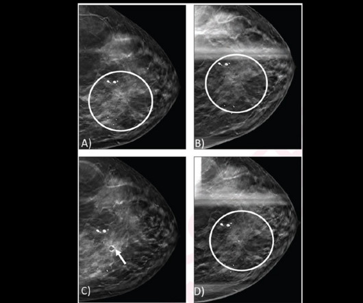
B) Spot craniocaudal view from subsequent diagnostic DBT mammogram shows persistence of architectural distortion (circle). D) Spot craniocaudal view from diagnostic mammogram performed 12 months later shows stable appearance of an architectural distortion (circle). No ultrasound correlate was identified (not shown).
He also discusses how this innovative solution is not just advancing diagnostic precision but also reshaping the way healthcare providers approach heart disease treatment and patientcare. Strange dives into how EchoSolv leverages cutting-edge technology to tackle one of cardiology's most challenging conditions.
We serve patients and physicians to improve diagnostics, treatments, and recovery. We are at the center of patientcare and we take that responsibility very seriously. Our mission of quality patientcare is achieved by adhering to high standards, improving processes, knowledge, skill, and the use of technology.
Description: This description provides an overview of a resource that explores how digital reporting is transforming the landscape of precision diagnostics in the realm of CT scans and MRI, enhancing the accuracy and efficiency of healthcare services.
Healthcare professionals use ultrasound machines for several purposes, including diagnostic exams, cardiac assessments, pregnancy evaluation, guiding procedures and numerous other areas of patientcare. However, the cost of an ultrasound machine can vary significantly depending on the type of machine and configuration.
This non-invasive procedure is ideal for patients who experience chest pains or heart conditions, offering an alternative to invasive diagnostic techniques. Detect Abdominal and Pelvic Conditions For patients with abdominal pain, gastrointestinal issues, or similar abnormalities, CT scans can help identify these issues.
MRI-Scan-Teleradiology In the vast and diverse canvas of India, where mountains, plains, and remote villages paint a challenging geographical tapestry, the triumph of teleradiology emerges as a revolutionary force in elevating patientcare.
The new Scope of Practice and Clinical Standards for the Diagnostic Medical Sonographer was released last month by the Society of Diagnostic Medical Sonographers. They are distinguished from contrast agents used with CT and MRI , which have less favorable safety profiles.
an innovative world leader delivering end-to-end products and solutions through a comprehensive portfolio inclusive of precision diagnostic imaging modalities, announces that on December 7, 2023 the European Commission (EC) has granted the Marketing Authorisation for Vueway ( gadopiclenol ) in the European Union (EU). mmol/kg gadobutrol 1,2.
The earlier breast cancer is detected through diagnostic imaging, the better chance there is for successful treatment with surgery, radiation therapy, or chemotherapy. Consistent mammography and diagnostic imaging appointments can help stop this number from continuing to rise. Our entire staff is committed to exceptional patientcare.
MRI-Scan-Teleradiology Description: This description provides an overview of a resource that explores how online reporting for CT scans and MRIs is ushering in a new era of seamless diagnostics, enhancing the efficiency and accessibility of radiological information in the digital age.
milla1cf Fri, 01/26/2024 - 21:11 January 26, 2024 — InkSpace Imaging , a leader in innovative diagnostic medical device technology, is proud to announce it received FDA clearance for its next-generation Small Body Array for the Siemens Healthineers Magnetom Skyra and Vida series 3T MRI scanners. Friday, January 26, 2024 - 21:11
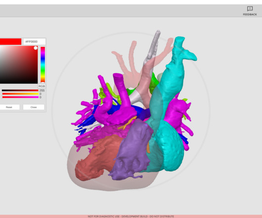
We like our surgeons to think and act at the speed of thought, and any friction created by complex software takes opportunities away from patientcare.” Radiologists and clinicians alike are turning to 3D imaging to better diagnose conditions and view anatomical structures. hospitals at no cost.
We organize all of the trending information in your field so you don't have to. Join 5,000 users and stay up to date on the latest articles your peers are reading.
You know about us, now we want to get to know you!
Let's personalize your content
Let's get even more personalized
We recognize your account from another site in our network, please click 'Send Email' below to continue with verifying your account and setting a password.


Let's personalize your content