This site uses cookies to improve your experience. To help us insure we adhere to various privacy regulations, please select your country/region of residence. If you do not select a country, we will assume you are from the United States. Select your Cookie Settings or view our Privacy Policy and Terms of Use.
Cookie Settings
Cookies and similar technologies are used on this website for proper function of the website, for tracking performance analytics and for marketing purposes. We and some of our third-party providers may use cookie data for various purposes. Please review the cookie settings below and choose your preference.
Used for the proper function of the website
Used for monitoring website traffic and interactions
Cookie Settings
Cookies and similar technologies are used on this website for proper function of the website, for tracking performance analytics and for marketing purposes. We and some of our third-party providers may use cookie data for various purposes. Please review the cookie settings below and choose your preference.
Strictly Necessary: Used for the proper function of the website
Performance/Analytics: Used for monitoring website traffic and interactions
This year’s trip along the Road to RSNA for digital x-ray features a few familiar mileposts – AI for chest x-ray studies, for instance – but notably also significant research into how technology and new techniques can reduce radiation exposure in patients. DAEC system reduces portable x-ray radiation doses Sunday, December 1 | 1:20 p.m.-1:30
More than 60% of diagnostic radiology and radiation therapy staff experience workplace violence, according to a study published January 9 in Radiography. Yet no systematic review has been published on the issue in medical radiation science, the authors noted.
. | M7-SSPH05-2 | Room N229 Findings will be presented in this Monday afternoon presentation on organ-specific ionizing radiation doses in neonatal patients who undergo interventional procedures for congenital heart disease (CHD). Gy-cm2, with organ-specific radiation doses highest for lung from frontal view (8.1 Louis, and colleagues.
12, 2025 Konica Minolta Healthcare Americas, has published a case study by clinicians in the pulmonary and radiology departments at ASST Fatebenefratelli Sacco (Milan, Italy) demonstrating the use of Dynamic Digital Radiography (DDR) to help definitively diagnose diaphragm dysfunction. tim.hodson Fri, 02/14/2025 - 15:14 Feb.12,
The American Society of Radiologic Technologists (ASRT) said it supports a new Pennsylvania bill that requires licensing of medical imaging professionals, radiation therapists, radiologist assistants, and trainees. The bill also addresses the field of radiologist assistants, which are advanced radiography specialists.
He earned his medical degree at the University of Alabama School of Medicine and completed a residency in diagnostic radiology at the University of Florida. An assistant professor of radiology and radiological science, Francis Deng, MD, is the Johns Hopkins radiology department's co-director of medical student diagnostic radiology electives.
The company will showcase the clinical analysis of Canon’s Intelligent Noise Reduction (Intelligent NR) that provides superior image quality while lowering radiation dosing in pediatric digital radiography at the Radiological Society of North America Annual Meeting 2023 (RSNA) , McCormick Place Convention Center, Chicago, IL Nov.
In an open forum, Yi Xiang Tay, of Singapore University Hospital's radiography and diagnostic imaging department, shared his team's research. In his presentation, Tay said the combination reduced radiation from CTs and x-rays and reduced unnecessary intravenous contrast administration for patients, mitigating side-effects of contrast.
Chest dynamic digital radiography (DDR) may have received a boost toward clinical use in patients with lung disorders, with researchers developing AI to perform time-consuming analysis involved in the technology, according to researchers in New York City. The study was published March 29 in Chest Pulmonary.
Led by Eli Atar, MD, director of the department of imaging, and Ahuva Grubstein, MD, department of diagnostic radiology, the study will assess the diagnostic capabilities of the Nanox.ARC’s tomographic imaging system compared with conventional 2D radiography for detecting lung and chest disease in adults.
Radiography has exploded into a variety of modalities and specialisms from CT to Ultrasound to MRI; all driven by research and development. I’m also passionate about education, research, and (of course) radiography. I also provide support to all trials requiring access to our radiography department, regardless of the imaging modality.
Positioning Techniques Importance of Proper Positioning Correct positioning is crucial for obtaining clear, diagnostic-quality images. Poor positioning can lead to retakes, increased radiation exposure, and misdiagnoses. Diagnostic Imaging Systems has wide variety of patient positioners to aid veterinary technicians.
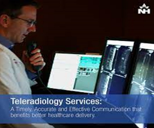
Teleradiology Introduction: Dental radiography plays a pivotal role in modern dentistry, enabling practitioners to diagnose and treat various oral health conditions. However, the field of dental radiography has undergone significant advancements, along with its own set of challenges and triumphs.
The ability to lower radiation doses without a loss in image quality also has considerable benefits in neonatal and pediatric imaging where imaging at the lowest possible dose is critical. In healthcare, it has the potential to improve diagnostic and treatment processes.
MRI-Scan-Teleradiology Introduction: Dental radiography is an essential component of modern dentistry, offering valuable insights into oral health and guiding treatments with precision. Patient-Centered Approach: Dental radiography starts with the patient. Let’s explore the key aspects of this intricate process.
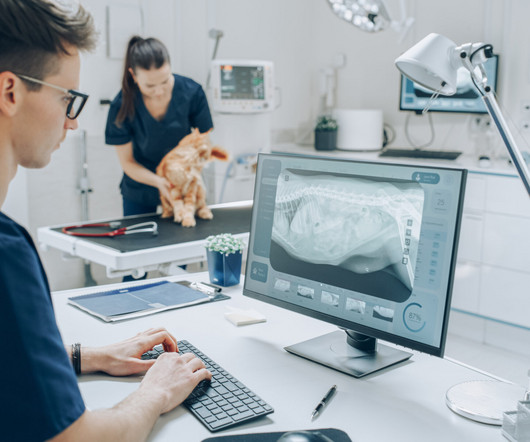
Introduction : In the past decade, veterinary medicine has witnessed a transformative shift with the adoption of digital radiography systems in place of traditional film-based methods. This transition promises a myriad of benefits, ranging from improved image quality and diagnostic accuracy to enhanced workflow efficiency.
Dynamic chest radiography (DCR) shows potential as a tool to investigate lung health in people with cystic fibrosis (CF), according to research published February 13 in Clinical Radiology. and colleagues. Yet FEV1 may not always reflect the severity of the airway obstruction, they noted. The full study is available here.
Teleradiology-in-Flat-World Introduction: Radiography in dentistry is a dynamic field with a myriad of applications and continuous innovations. This blog navigates through the world of dental radiography, highlighting its diverse applications and the ongoing advancements that shape the future of oral health.
X-ray radiography is a noninvasive diagnostic method that uses X-rays—electromagnetic radiation—to produce images of the body's internal structures. Essential in medical fields for diagnosing injuries and diseases and monitoring treatment progress, X-ray radiography is a critical tool in patient care and medical decision-making.
Introduction: The world of radiography is one filled with immense responsibility and precision, where the journey to capture images of the human body’s inner workings is not without its challenges. Radiation Safety: A Top Priority: Radiation safety is paramount in radiography.
This was the case for diagnostic purposes but also for follow-up imaging at 3, 6 and 12 months after re-alignment procedures, fusions or arthroplasties. Cone beam CT, be it weight bearing or not, is equivalent to 3D radiography and that is where most of the healthcare benefits are for patients. I use WBCT every day.
Teleradiology-India Introduction: “Capturing the Invisible” takes you on a captivating journey into the world of X-rays, unveiling their impactful role in diagnostics. How X-rays are generated, interact with the human body, and create diagnostic images. The pivotal role of X-rays in early detection and accurate diagnosis.
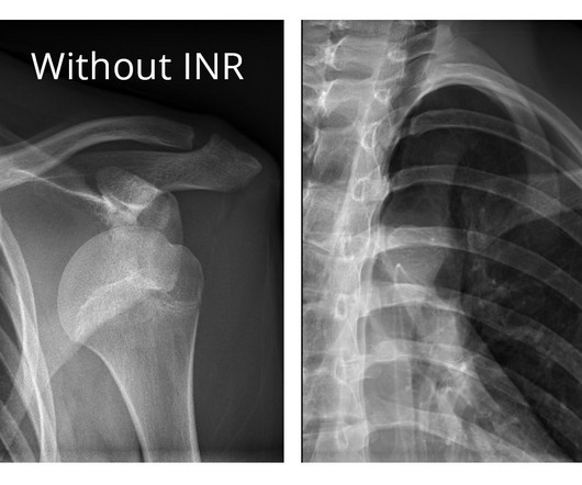
Generally limited on tissue differentiation, portable chest radiography can be ineffective at accurately spotting complex pulmonary issues and sometimes even to localize the tips of lines and tubes. Our radiographic spectral images separate materials such as water (i.e., soft tissue, lung lesions etc.) and calcium (i.e.
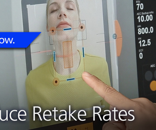
For example, IEC Exposure Index allows quick assessment of the amount of radiation used to create the image; while Deviation Index immediately compares the chosen exposure to your facility’s specific target goal. The result: image quality comparable to images acquired with an anti-scatter grid but at a lower radiation dose.
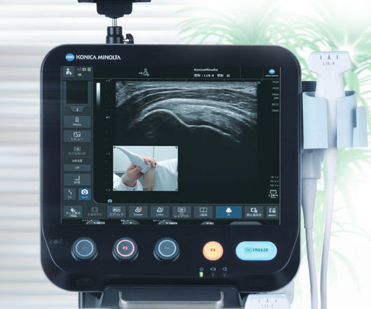
announced today several new solutions in digital radiography (DR) and ultrasound that will be introduced at the 2023 Radiology Society of North America (RSNA) Scientific Assembly and Annual Meeting from November 26-30 in Chicago, IL. This capability simplifies clinical workflow and reduces patient exposure to radiation dose.
About Shamie Kumar Shamie Kumar Shamie Kumar is a practicing HCPC Diagnostic Radiographer; graduated from City University London with a BSc Honors in DiagnosticRadiography in 2009 and is a part of Society of Radiographers with over 12 years of clinical knowledge and skills within all aspects of radiography. Coakley, Y.
This journey takes us from the early days of X-ray discovery by Wilhelm Roentgen to the cutting-edge digital and computational innovations that shape the modern landscape of diagnostic imaging. Chapter 2: The Art and Science of Radiography A closer look at the development of radiography, the first X-ray imaging method.
Radiography soon became a vital tool in medicine, and for decades, it relied on photographic film to capture and develop X-ray images. The Birth of Digital Radiography: The 21st century brought a seismic shift as X-ray imaging transitioned from film to digital radiography.
From Film to Digital: The transition from film-based X-ray systems to digital technology marked a turning point in dental radiography. Digital sensors and intraoral cameras offer instant image acquisition, reduced radiation exposure, and the ability to enhance and manipulate images for better diagnostics.
How X-rays are generated, interact with the human body, and create diagnostic images. Chapter 3: The Evolution of Radiography: From Shadows to Insights An exploration of the development of radiography, the earliest X-ray imaging technique. How these modalities expand diagnostic capabilities for specific medical conditions.
Benefits of Teleradiology to Telehealth Introduction: “Behind the Beams” is an intriguing journey that delves into the intricate world of X-ray technology, uncovering the perfect fusion of art and science that powers this essential diagnostic tool. How X-rays are generated, interact with matter, and produce diagnostic images.
In medical imaging and diagnostics, advanced image recognition has profound applications in medical imaging and diagnostics. Additionally, patients deserve the best possible diagnostic imaging as quickly as possible so they can get the proper diagnosis and begin the right treatment promptly.
Chest radiography is a common diagnostic tool, but significant training and experience is required to interpret exams correctly,” said lead researcher Louis L. AI tools can assist radiologists in interpreting chest X-rays , but their real-life diagnostic accuracy remains unclear.” Plesner, M.D., resident radiologist and Ph.D.
Not only are children more radiosensitive than adults (the cancer risk per unit dose of ionizing radiation is higher), but children also have a longer expected lifetime, which puts them at greater risk of cancer following radiation exposure.(1) The balance of dose and image quality is even more important in pediatric medical imaging.
In this exploration, we will weave together the history, science, and practical applications of X-ray imaging, providing a holistic understanding of this invaluable diagnostic tool. How X-rays are generated, interact with human tissue, and create diagnostic images. Real-life case studies illustrating the diagnostic power of X-rays.
From its discovery by Wilhelm Roentgen to modern applications, we will illuminate the shadows to provide a deep understanding of this essential diagnostic tool. The historical context of Wilhelm Roentgen’s discovery and the birth of this revolutionary diagnostic tool. How each modality is used for different clinical purposes.
Common Indications Scaphoid Fracture A feasible alternative to MDCT for the detection of extremity fractures at a reduced radiation dose. Patients with metal implants can obtain advanced diagnostic imaging. The use of cone-beam computed tomography (CBCT) in radiocarpal fractures: a diagnostic test accuracy meta-analysis.
To meet that goal, we had to get the best possible equipment and that’s why we turned to Fujifilm Healthcare Americas Corporation for the very latest in diagnostic imaging systems.” The recent investment in a full portfolio of diagnostic imaging equipment is the latest testament to that philosophy.
4) Precise patient positioning in radiology is essential to obtaining accurate diagnostic information to aid in effective patient care and for reducing a patient’s X-ray exposure due to retakes. 2 Unified Database for Rejected Image Analysis Across Multiple Vendors in Radiography.
Their expertise and skill in operating X-ray machines and positioning patients are crucial in obtaining clear and diagnostically valuable images. The Dance of Radiation Safety: Protecting Patients and Themselves: Radiation safety is a core concern for X-ray technicians. X-ray technicians are not only masters of technology.
The Birth of X-ray Technology: At the end of the 19th century, Wilhelm Conrad Roentgen’s discovery of X-rays opened up new possibilities in medical diagnostics. Traditional film-based X-rays gave way to digital radiography (DR) and computed radiography (CR).
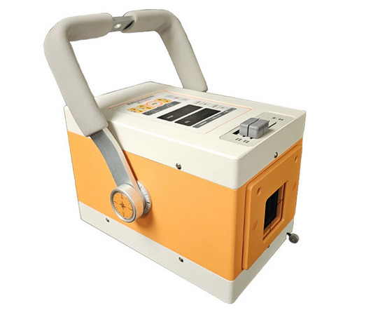
In the ever-evolving landscape of veterinary medicine, technological advancements continue to play a pivotal role in enhancing patient care and diagnostic capabilities. Radiation safety is a paramount concern in veterinary medicine, and portable x-ray units address this by incorporating features to minimize exposure.
Pelc added, “The AIxSCAN tomosynthesis X-ray scanner does not completely avoid the superimposition limitations of radiography, but the early results from the prototype system are very encouraging and suggest the technology can play a significant role. Advisory Board member, commenting on the ARC60 imaging platform capabilities.
We organize all of the trending information in your field so you don't have to. Join 5,000 users and stay up to date on the latest articles your peers are reading.
You know about us, now we want to get to know you!
Let's personalize your content
Let's get even more personalized
We recognize your account from another site in our network, please click 'Send Email' below to continue with verifying your account and setting a password.


Let's personalize your content