This site uses cookies to improve your experience. To help us insure we adhere to various privacy regulations, please select your country/region of residence. If you do not select a country, we will assume you are from the United States. Select your Cookie Settings or view our Privacy Policy and Terms of Use.
Cookie Settings
Cookies and similar technologies are used on this website for proper function of the website, for tracking performance analytics and for marketing purposes. We and some of our third-party providers may use cookie data for various purposes. Please review the cookie settings below and choose your preference.
Used for the proper function of the website
Used for monitoring website traffic and interactions
Cookie Settings
Cookies and similar technologies are used on this website for proper function of the website, for tracking performance analytics and for marketing purposes. We and some of our third-party providers may use cookie data for various purposes. Please review the cookie settings below and choose your preference.
Strictly Necessary: Used for the proper function of the website
Performance/Analytics: Used for monitoring website traffic and interactions
Intelligent virtual and AI-based collimation features appear to save radiographers time during x-ray image acquisitions – a key function for enabling more patient-focused workflows, according to a recent study. The study was published online May 18 in Radiography. Finally, VC was used to collimate in 2.4%
12, 2025 Konica Minolta Healthcare Americas, has published a case study by clinicians in the pulmonary and radiology departments at ASST Fatebenefratelli Sacco (Milan, Italy) demonstrating the use of Dynamic Digital Radiography (DDR) to help definitively diagnose diaphragm dysfunction. tim.hodson Fri, 02/14/2025 - 15:14 Feb.12,
ChatGPT-4 outperformed human clinicians in determining pretest and post-test disease probability after a negative test result involving chest radiographs and mammograms, according to a research letter published December 11 in JAMA Network Open.
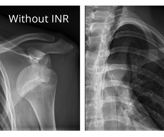
The company will showcase the clinical analysis of Canon’s Intelligent Noise Reduction (Intelligent NR) that provides superior image quality while lowering radiation dosing in pediatric digital radiography at the Radiological Society of North America Annual Meeting 2023 (RSNA) , McCormick Place Convention Center, Chicago, IL Nov.
He earned his medical degree at the University of Alabama School of Medicine and completed a residency in diagnostic radiology at the University of Florida. An assistant professor of radiology and radiological science, Francis Deng, MD, is the Johns Hopkins radiology department's co-director of medical student diagnostic radiology electives.
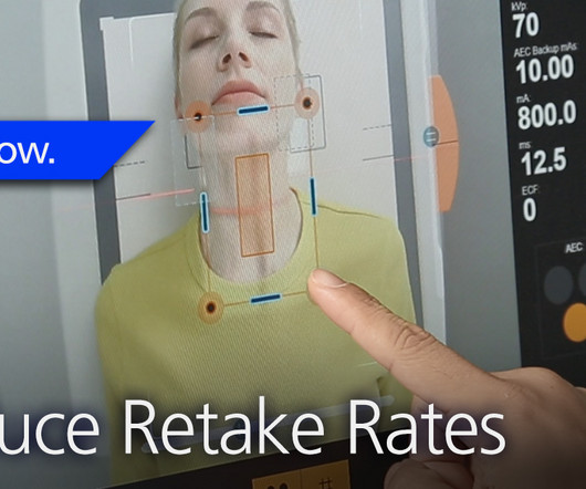
At RSNA 2023, look for AI-driven systems that radiographers can use to help make patient positioning faster and more precise, and bring consistency to the process, all of which help improve image quality and reduce the need for retakes. Even the most skilled radiographers can fail to get positioning just right.
In an open forum, Yi Xiang Tay, of Singapore University Hospital's radiography and diagnostic imaging department, shared his team's research. It was the endnote of a series of sessions focused on optimizing radiology services.
Dynamic digital radiography (DDR) has shown for the first time that it can be used to automatically capture lung signal changes during forced breathing in patients with chronic obstructive pulmonary disease (COPD), according to a recent study. and Japanese developers wrote. The subject is a 77-year-old male with VC of 1.68 L and FEV1 of 0.68
Chest dynamic digital radiography (DDR) may have received a boost toward clinical use in patients with lung disorders, with researchers developing AI to perform time-consuming analysis involved in the technology, according to researchers in New York City. a) Raw example of a dynamic digital radiograph. (b)
This ensures an unobstructed qualitative evaluation, and enables a quantitative evaluation of dark-field radiographs,” Urban et al wrote. Dark-field chest x-ray has just recently been translated to the clinical stage and its diagnostic value is currently being investigated with a clinical prototype, the authors wrote.
milla1cf Wed, 06/12/2024 - 21:59 June 12, 2024 — Carestream launched its Image Suite MR 10 Software to help deliver a boost to productivity and efficiency while enabling a more user-friendly imaging experience for radiographers. Image Suite MR 10 helps push the envelope further for even more focus on patient care.”
milla1cf Fri, 07/07/2023 - 21:32 July 7, 2023 — FUJIFILM Healthcare Americas Corporation, a leading provider of diagnostic and enterprise imaging solutions, today announced the U.S. launch of its latest addition to its advanced digital radiography suites, the D-EVO Suite OTCx.
Shamie Kumar describes how AI fits into a radiology clinical workflow and her perspective on how a clinical radiographer could use this to learn from and enhance their skills. If the AI findings are seen in PACS, how many radiographers actually log into PACS after taking a scan or X-ray? Can Radiographers Up-Skill?
11, 2025 Harrison.ai, a developer of AI-powered medical diagnostic support and workflow solutions, has announced the accelerated expansion of its operations into the United States a move supported by US$112 million of Series C funding. Radiologists using Harrison.ai's technology have seen an over 45% increase in diagnostic accuracy1.
Kim Mason Kim Mason, an Audit and Research Radiographer for Mid Yorkshire Teaching Hospitals Trust, talks about their role as well as the value of radiographer engagement in research activities and how to get involved. Hi, I’m Kim and I am an alternative-styled, funky-haired, septum-pierced, disabled Audit and Research Radiographer.
Central Vein Sign in Multiple Sclerosis: A Comparison Study of the Diagnostic Performance of 3T versus 7T MRI. Commercially Available Chest Radiograph AI Tools for Detecting Airspace Disease, Pneumothorax, and Pleural Effusion. Generative Artificial Intelligence for Chest Radiograph Interpretation in the Emergency Department.
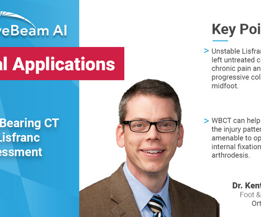
Whenever bilateral standing radiographs would have been needed, a WBCT was performed instead. This was the case for diagnostic purposes but also for follow-up imaging at 3, 6 and 12 months after re-alignment procedures, fusions or arthroplasties. I use WBCT every day. It is also the most time and cost-efficient modality.
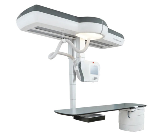
Enabling radiographic and low-dose fluoroscopy exams in the same room, the Adora DRFi system emphasizes a variety of advances for flexibility with patient positioning.
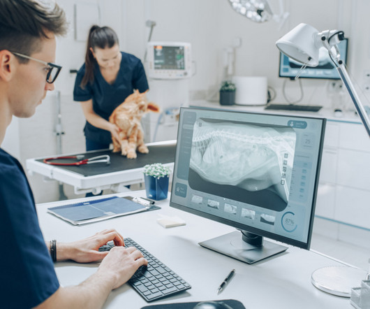
Veterinary technicians play a pivotal role in the radiographic process within veterinary practices. Positioning Techniques Importance of Proper Positioning Correct positioning is crucial for obtaining clear, diagnostic-quality images. Diagnostic Imaging Systems has wide variety of patient positioners to aid veterinary technicians.
Dynamic chest radiography (DCR) shows potential as a tool to investigate lung health in people with cystic fibrosis (CF), according to research published February 13 in Clinical Radiology. and colleagues. Yet FEV1 may not always reflect the severity of the airway obstruction, they noted. The full study is available here.
to demonstrate portable digital radiography and its effectiveness at high altitudes. Cairnie says, “Radiograph technology is critical for diagnosing several conditions, including bone fracture and chest conditions such as Tuberculosis.
Our hope is that we'll be able to determine how to use this as a diagnostic tool in actual practice,” he said. He added that by the age of 50 or 60, many or most people have already had a CT scan for diagnostic purposes. “We We can first take advantage of all those scans,” he said.
Teleradiology-in-Flat-World Introduction: Radiography in dentistry is a dynamic field with a myriad of applications and continuous innovations. This blog navigates through the world of dental radiography, highlighting its diverse applications and the ongoing advancements that shape the future of oral health.
MRI-Scan-Teleradiology Introduction: Dental radiography is an essential component of modern dentistry, offering valuable insights into oral health and guiding treatments with precision. Patient-Centered Approach: Dental radiography starts with the patient. Quality Assurance: Dental radiographers are meticulous in quality assurance.
Repeating imaging exams increases the workload of your radiographers who are already stretched too thin; increases the exposure of the affected patients; and contributes to patients’ reduced confidence and satisfaction with your imaging department. The Audio Assist makes it easier for radiographers to hear the patients.
In medical imaging and diagnostics, advanced image recognition has profound applications in medical imaging and diagnostics. AI tools that enable radiographers to separate noise from an image already exist today. Carestream’s Smart Assist features can help you ease the burden on your radiographers.
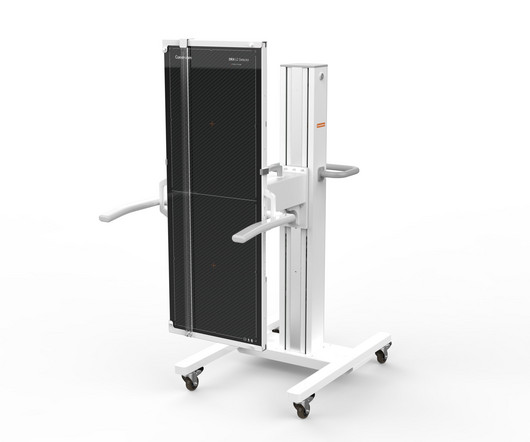
milla1cf Thu, 05/18/2023 - 15:04 May 18, 2023 — Carestream Health’s new, versatile DRX-LC Detector is designed to improve patient comfort, image quality and diagnostic confidence, and productivity for long-length image capture in orthopedics. The second-generation long-length detector has several new features.
Generally limited on tissue differentiation, portable chest radiography can be ineffective at accurately spotting complex pulmonary issues and sometimes even to localize the tips of lines and tubes. Our radiographic spectral images separate materials such as water (i.e., soft tissue, lung lesions etc.) and calcium (i.e.
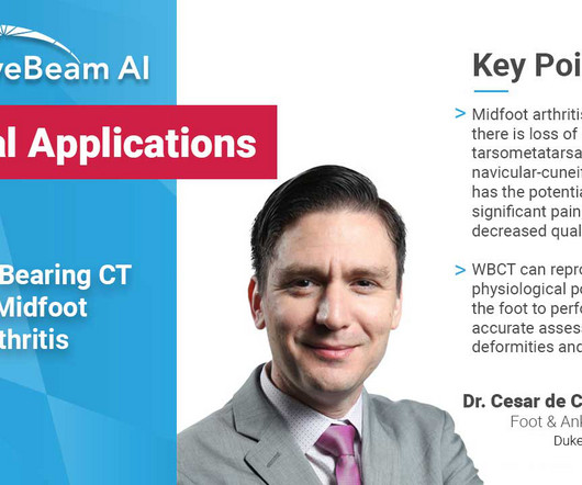
Key Points: Currently plain radiographs are the standard method in diagnosing syndesmotic ankle injuries even though the distal tibiofibular joint cannot be assessed due to superposition of the osseous structures in the foot.
Teleradiology Introduction: The landscape of chest radiography is undergoing a revolutionary transformation, with deep learning at its forefront. This blog post explores how deep learning algorithms are reshaping the future of chest radiography, particularly in the realm of pneumonia detection and classification.
Again, while radiographers and radiologists are capturing and diagnosing images, the people signing off on the actual financial investment are the administrators, the CEOs, clinical engineering leaders, product committee chairs, and chief medical officers. Radiologists want to know how much more accurate they will be with their diagnostics.
“An advanced DR mobile imaging system can dramatically improve patient care, diagnostic confidence and mobile imaging throughput, but the cost of the equipment has been out of reach for some facilities,” said Jordan Berry, Global Marketing Manager, Carestream Health.
milla1cf Thu, 11/23/2023 - 06:00 November 23, 2023 — Fujifilm Healthcare Americas Corporation, a leading provider of diagnostic and enterprise imaging solutions, is unveiling several new medical systems at the 2023 Radiological Society of North America ( RSNA ) annual meeting, booth #1929, held November 26 – 30 at McCormick Place in Chicago.
A fundamental goal of radiographers is to complete an imaging exam that provides sufficient information for an accurate clinical diagnosis–and at the lowest possible dose. We also have a Detector Verification alert that signals radiographers when they choose the wrong detector–which would result in the need to repeat the exam.
In the third blog of her series on AI and the radiographer, Shamie Kumar explores the impact on the radiographer when AI is integrated within an imaging modality. The question to explore in this blog is when AI is integrated within an imaging modality itself and how that may impact a radiographer.
But how will AI in the workplace affect the radiographer and how does it differ from the red dot system radiographers are so familiar with? The Red Dot System Often one of the first courses a newly qualified radiographer attends is the red dot course. What does AI do that a radiographer doesn’t already?
Building on its century-long foundation of imaging research science, the company will showcase its diagnostic imaging solutions that help improve clinical outcomes, enhance the imaging experience for users and patients, and strengthen the financial position of healthcare facilities. “The
X-rays, a cornerstone of medical diagnostics for over a century, remain crucial in today's healthcare landscape. This radiographic technique provides vital insights into the body's internal structure, assisting in accurate diagnosis and treatment planning. But how exactly are X-rays performed?
Diagnosis The diagnosis of Lisfranc injuries may be challenging on plain radiographs alone. Radiographs were indeterminate. Comparative assessment of midfoot osteoarthritis diagnostic sensitivity using weightbearing computed tomography vs weightbearing plain radiography. Accurately assess healing of TMT joint fusions 5.
In addition, WBCT better quantifies 3 the structural deformity of Chopart, talonavicular, and calcaneocuboid joints when compared to conventional radiography and non-weight bearing computed tomography images. However, the severity of deformity and involved joints were difficult to determine on plain radiographs alone. Eur J Radiol.
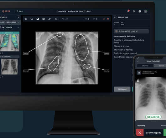
Qure’s chest X-ray based qXR-LN uses artificial intelligence to identify and localize lung nodules, marking another significant milestone for the organization, strengthening its standing as a pioneer in the realm of AI-powered advancements for plain film radiography and medical imaging. stands at the forefront of diagnostic advancement.
Radiography training in many institutions around the world is heavily process-driven; that is, radiographers are taught the technical requirements of a CT scan and learn the clinical side on the job.
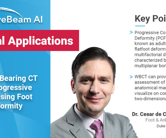
A weight bearing CT scan can: Provide an assessment of important anatomical markers of pronounced hindfoot deformity and peritalar subluxation (PTS), difficult to visualize on conventional two-dimensional radiographs 1. Allow for accurate evaluation of subtalar joint subluxation as well as sinus tarsi and subfibular impingement 2 .
Powered by Eclipse, ImageView utilizes AI and Carestream’s proprietary algorithms to provide our most advanced image processing capabilities for superb image quality and diagnostic confidence. Physicians have better diagnostic confidence when they can view radiographs in the manner most suitable to their preferences.
We organize all of the trending information in your field so you don't have to. Join 5,000 users and stay up to date on the latest articles your peers are reading.
You know about us, now we want to get to know you!
Let's personalize your content
Let's get even more personalized
We recognize your account from another site in our network, please click 'Send Email' below to continue with verifying your account and setting a password.


Let's personalize your content