This site uses cookies to improve your experience. To help us insure we adhere to various privacy regulations, please select your country/region of residence. If you do not select a country, we will assume you are from the United States. Select your Cookie Settings or view our Privacy Policy and Terms of Use.
Cookie Settings
Cookies and similar technologies are used on this website for proper function of the website, for tracking performance analytics and for marketing purposes. We and some of our third-party providers may use cookie data for various purposes. Please review the cookie settings below and choose your preference.
Used for the proper function of the website
Used for monitoring website traffic and interactions
Cookie Settings
Cookies and similar technologies are used on this website for proper function of the website, for tracking performance analytics and for marketing purposes. We and some of our third-party providers may use cookie data for various purposes. Please review the cookie settings below and choose your preference.
Strictly Necessary: Used for the proper function of the website
Performance/Analytics: Used for monitoring website traffic and interactions
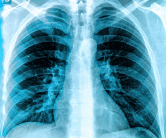
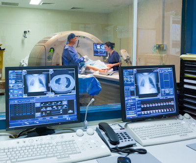
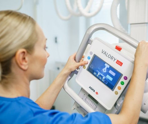
Intelligent virtual and AI-based collimation features appear to save radiographers time during x-ray image acquisitions – a key function for enabling more patient-focused workflows, according to a recent study. ATC and SVO enable the radiographer to save time during chest or stitched examinations.
Plain hip x-rays may help screen patients for osteoporosis before they undergo total hip arthroplasty (THA), according to a study published November 14 in Scientific Reports. However, several studies have suggested that indices on plain hip x-rays may be reliable screening tools in female or Asian ethnicities.
German developers of dark-field chest x-ray appear to have overcome a technical limitation of the technology – namely, adjusting for photon scattering caused by interferometers used in the experimental system. To overcome this limitation, the researchers developed a deconvolution-based correction method for the induced artifacts.
Reading Time: 10 minutes read By Henry Williams, Carestream Area Vice President, Sales Western Nowadays, with hospital budgetary restrictions at the forefront of the purchasing decision making process, it seems like the X-Ray market, like everything else, is not immune to the current state of the economy. But is that really the case?
In a study described as a “competition between radiologists,” participants tasked with identifying abnormal findings on chest x-rays performed better with AI assistance than without AI assistance – though not by much and not in all cases, according to a group in Nanjing, Jiangsu, China.
Diagnostic imaging tests are tools used by physicians to diagnose a range of medical conditions. For X-rays, it usually takes less than 10 minutes. Why Diagnostic Imaging Methods Are Important Medical imaging is used to diagnose, monitor, and treat medical problems. It can take up to 90 minutes, max.
Common diagnostic tests for pulmonary disorders include chest x-rays and pulmonary function tests (PFTs). a) Raw example of a dynamic digital radiograph. (b) The digital technology limits radiation exposure to patients compared with standard chest x-rays, they wrote.
DDR is a novel functional imaging technique that uses sequential images obtained by a pulsed x-ray generator and a flat panel detector with a large field of view. frames per second (COPD patients) or 15 frames per second (healthy volunteers) with synchronized pulsed x-rays. Dynamic image data was acquired at 7.5
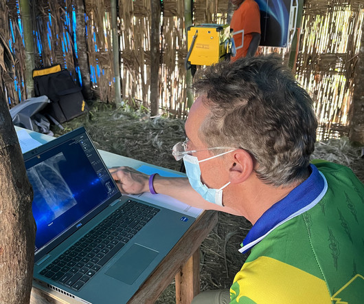
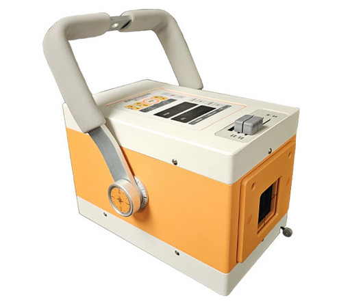
milla1cf Mon, 04/01/2024 - 11:44 April 1, 2024 — MinXray , a leading manufacturer of imaging systems for medical and veterinary use, recently sent its Impact Wireless X-ray system with a group of researchers and medical personnel to the YUS Conservation Area in Papua New Guinea.
Substituting less energy-consuming ultrasound for x-ray or CT reduced energy use by as much as 8% during diagnostic radiology processes and 31.2% for total CFCs, while demonstrating an increase in diagnostic activity of 11.8%, Masperi said. Stage two consisted of a retrospective review of exams performed and their timing.
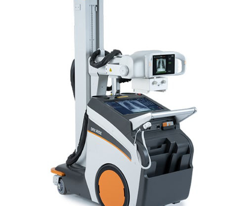
milla1cf Wed, 05/10/2023 - 22:58 May 10, 2023 — Expanding its industry-leading mobile X-ray portfolio, Carestream Health has introduced the DRX-Rise Mobile X-ray System. The system’s two touchscreen displays provide two work zones to accelerate productivity further.
In an open forum, Yi Xiang Tay, of Singapore University Hospital's radiography and diagnostic imaging department, shared his team's research. In his presentation, Tay said the combination reduced radiation from CTs and x-rays and reduced unnecessary intravenous contrast administration for patients, mitigating side-effects of contrast.
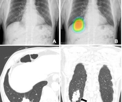
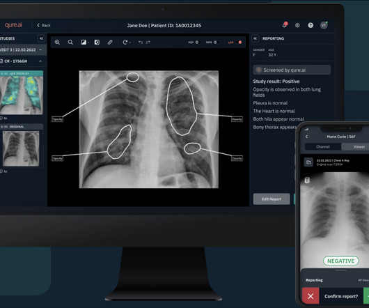
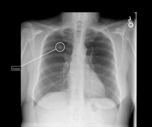
Qure’s chest X-ray based qXR-LN uses artificial intelligence to identify and localize lung nodules, marking another significant milestone for the organization, strengthening its standing as a pioneer in the realm of AI-powered advancements for plain film radiography and medical imaging.
Central Vein Sign in Multiple Sclerosis: A Comparison Study of the Diagnostic Performance of 3T versus 7T MRI. Commercially Available Chest Radiograph AI Tools for Detecting Airspace Disease, Pneumothorax, and Pleural Effusion. Generative Artificial Intelligence for Chest Radiograph Interpretation in the Emergency Department.
How would you rate the value you are getting from your current X-ray imaging equipment? Powered by Eclipse, ImageView utilizes AI and Carestream’s proprietary algorithms to provide our most advanced image processing capabilities for superb image quality and diagnostic confidence.
The study, published in the journal Diagnostics, highlights the clinical value of DDR through its unique ability to evaluate diaphragm movement in real time and integrate dynamic functional information with static anatomical data to provide a quantitative assessment of diaphragmatic movement, including excursion and speed.
At RSNA 2023, look for AI-driven systems that radiographers can use to help make patient positioning faster and more precise, and bring consistency to the process, all of which help improve image quality and reduce the need for retakes. Even the most skilled radiographers can fail to get positioning just right.
. | R1-SSCH09-5 | Room E352 An AI algorithm can find radiographic markers for osteoporosis that are common but often not reported on radiology reports, according to this scientific paper. The dataset consisted of 519 chest x-rays from patients ages 65 and older, collected from outpatient clinics.
a key global supplier of portable, compact digital imaging equipment, has earned a Guinness World Record for the “ highest altitude operating an X-ray machine ” while on an expedition with both MinXray and artificial intelligence company Qure.ai. to demonstrate portable digital radiography and its effectiveness at high altitudes.
In addition, an in-person demonstration called Radiology Reimagined: AI, innovation, and interoperability in practice is designed to showcase new technologies and communications standards needed to integrate AI into the diagnostic workflow, according to the RSNA. 10:50 a.m. | 8:10 a.m. | 8:40 a.m. | 1:40 p.m. | 1:50 p.m. | 8:30 a.m. |
11, 2025 Harrison.ai, a developer of AI-powered medical diagnostic support and workflow solutions, has announced the accelerated expansion of its operations into the United States a move supported by US$112 million of Series C funding. Radiologists using Harrison.ai's technology have seen an over 45% increase in diagnostic accuracy1.
milla1cf Fri, 07/07/2023 - 21:32 July 7, 2023 — FUJIFILM Healthcare Americas Corporation, a leading provider of diagnostic and enterprise imaging solutions, today announced the U.S. X-rays are the most widely used diagnostic tests, accounting for 60% of all imaging studies conducted.
Teleradiology-India Introduction: “X-ray Visionaries” takes you on a compelling journey to unveil the expertise of radiographers and technologists, the unsung heroes of X-ray technology. The importance of minimizing radiation exposure while ensuring precision in diagnostic imaging.
The studies include two oral presentations and five posters, and address AI advancements for chest x-ray reporting and breast cancer risk assessment, the company said.
Spirometry, specifically forced expiratory volume of air in 1 second (FEV1), is a prognostic marker in cystic fibrosis, and alongside chest x-ray findings is the primary method for assessing lung health in patients. Yet FEV1 may not always reflect the severity of the airway obstruction, they noted. The full study is available here.
The other role could be that of a limited x-ray machine operator. The department of radiology teaches a diagnostic technologies in healthcare course, principally around imaging technologies, for which 3,000 people have signed up, Rubin said. "It
The authors note that ionizing radiation is the basis for the production of diagnosticX-rays, however it has long been proven to increase the risk of cancer. 26 –29, 2023 in North Hall, Level 3, booth #7913.
Additionally, Carestream will display: Carestream’s full line of DR detectors — state-of-the-art DR detector options that are available in a variety of sizes to fit a facility’s workflow and budget, including the award-winning, glass-free Lux 35 Detector and small-format DRX Plus 2530C Detector.
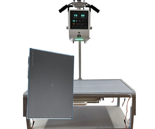
A fundamental goal of radiographers is to complete an imaging exam that provides sufficient information for an accurate clinical diagnosis–and at the lowest possible dose. To make it easier for readers, I’ve organized the available solutions into three exam types: general X-ray, chest imaging, and pediatric imaging.
Shamie Kumar describes how AI fits into a radiology clinical workflow and her perspective on how a clinical radiographer could use this to learn from and enhance their skills. If the AI findings are seen in PACS, how many radiographers actually log into PACS after taking a scan or X-ray? Can Radiographers Up-Skill?
In veterinary medicine, having access to reliable diagnostic tools is essential for providing high-quality care to your patients. X-ray systems are a fundamental part of this, but the choice between a mobile X-ray system and a stationary one can greatly impact the workflow and efficiency of your clinic.
Closeup of X-ray photography of human brain Introduction: In the world of modern medicine, there exists a fascinating blend of art and science, where the careful use of technology and technique converges to reveal the hidden truths within the human body. Radiographic film, once the primary medium, has given way to digital sensors.
13, 2024 — Agfa Radiology Solutions will feature live demonstrations of state-of-the-art digital X-ray rooms, mobile imaging solutions and software in its booth (#2565) at RSNA 2024 in Chicago in December. The Agfa booth also will feature an interactive touch screen kiosk on which visitors can compare radiographs. 3, 3-5 p.m.,
Introduction: Delve into the illuminating world of X-rays and their pivotal role in diagnosing and monitoring Inflammatory Bowel Disease (IBD). This exploration sheds light on the radiological insights that X-ray imaging provides, offering valuable information for the comprehensive understanding and management of IBD. **1.
The Reveal Mobi Pro integrates KA Imaging’s Reveal 35C detector with SpectralDR technology into a complete mobile X-ray solution. The Reveal 35C detector mimics the workflow, dose, and techniques of state-of-the-art mobile DR X-ray. Our radiographic spectral images separate materials such as water (i.e.,
In the ever-evolving landscape of veterinary medicine, technological advancements continue to play a pivotal role in enhancing patient care and diagnostic capabilities. One such innovation that has transformed the way veterinarians approach imaging is the evolution of portable x-ray units.
X-rays, a cornerstone of medical diagnostics for over a century, remain crucial in today's healthcare landscape. This radiographic technique provides vital insights into the body's internal structure, assisting in accurate diagnosis and treatment planning. But how exactly are X-rays performed?
Agfa’s comprehensive portfolio supports “The Next Generation” in medical imaging by using intelligent and innovative technologies to ensure every X-ray image counts. Our productivity features and ‘one image is all it takes’ approach empower each X-ray expert to work more efficiently.
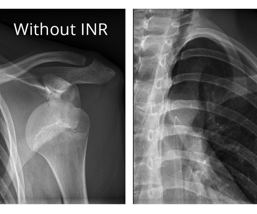
milla1cf Thu, 05/18/2023 - 15:04 May 18, 2023 — Carestream Health’s new, versatile DRX-LC Detector is designed to improve patient comfort, image quality and diagnostic confidence, and productivity for long-length image capture in orthopedics. The second-generation long-length detector has several new features.
Introduction: Dental X-ray technologists, also known as dental radiographers, are the unsung heroes behind the scenes of every successful dental diagnosis and treatment. In this blog, we’ll take you on a journey inside the jaw and explore a day in the life of a dental X-ray technologist.
Tailored for incidental findings on chest radiographs, the qXR for Lung Nodule (qXR-LN) software utilizes artificial intelligence (AI) to help detect suspected pulmonary nodules ranging between 6 to 30 mm.
We also are showcasing our expanding line of X-ray solutions that deliver high-quality imaging at a more affordable price.” These will include the DRX-Revolution Mobile X-ray System, Vita Flex CR with the DRYVIEW 5950 Laser Imager, and the DRX Plus Detectors including the Lux 35 Detector.
We organize all of the trending information in your field so you don't have to. Join 5,000 users and stay up to date on the latest articles your peers are reading.
You know about us, now we want to get to know you!
Let's personalize your content
Let's get even more personalized
We recognize your account from another site in our network, please click 'Send Email' below to continue with verifying your account and setting a password.


Let's personalize your content