This site uses cookies to improve your experience. To help us insure we adhere to various privacy regulations, please select your country/region of residence. If you do not select a country, we will assume you are from the United States. Select your Cookie Settings or view our Privacy Policy and Terms of Use.
Cookie Settings
Cookies and similar technologies are used on this website for proper function of the website, for tracking performance analytics and for marketing purposes. We and some of our third-party providers may use cookie data for various purposes. Please review the cookie settings below and choose your preference.
Used for the proper function of the website
Used for monitoring website traffic and interactions
Cookie Settings
Cookies and similar technologies are used on this website for proper function of the website, for tracking performance analytics and for marketing purposes. We and some of our third-party providers may use cookie data for various purposes. Please review the cookie settings below and choose your preference.
Strictly Necessary: Used for the proper function of the website
Performance/Analytics: Used for monitoring website traffic and interactions
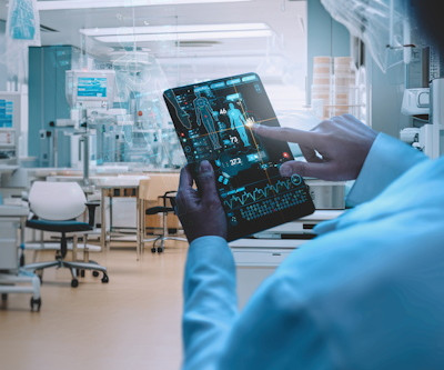
While radiographers are concerned about job security, they are also optimistic about AI’s role in their future workflows, according to a presentation given March 1 at ECR 2024. Radiographers appear optimistic about the future of radiographer job roles and responsibilities,” Walsh said. Of the total respondents, 31.3%
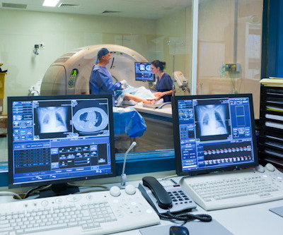
Intelligent virtual and AI-based collimation features appear to save radiographers time during x-ray image acquisitions – a key function for enabling more patient-focused workflows, according to a recent study. ATC and SVO enable the radiographer to save time during chest or stitched examinations. The full study is available here.
Some of my radiological heroes would report a staggering 30,000 to 40,000 radiographs a year. Some even [startled gasp] gave up reporting plain radiographs. Many diagnostic radiologists became pure CT and MRI specialists. I still don’t know how they did it. The neocortex rarely had to engage.
AI for thoracic imaging includes using it for reading chest radiographs and low-dose chest CT scans for lung cancer screening and for triaging pulmonary embolism on chest CT scans, the group noted. These LLMs offer opportunities ranging from generating text reports from images to explaining examination results to patients."
An easily accessible screening tool for osteopenia or osteoporosis using plain hip radiographs is of great importance for the orthopedic hip surgeons to avoid potential surgical risks while performing total hip arthroplasty due to osteopenia and osteoporosis,” noted lead author Sebastian Rohe, MD, and colleagues.
ChatGPT-4 outperformed human clinicians in determining pretest and post-test disease probability after a negative test result involving chest radiographs and mammograms, according to a research letter published December 11 in JAMA Network Open.
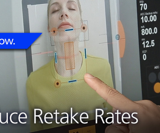
At RSNA 2023, look for AI-driven systems that radiographers can use to help make patient positioning faster and more precise, and bring consistency to the process, all of which help improve image quality and reduce the need for retakes. Even the most skilled radiographers can fail to get positioning just right.
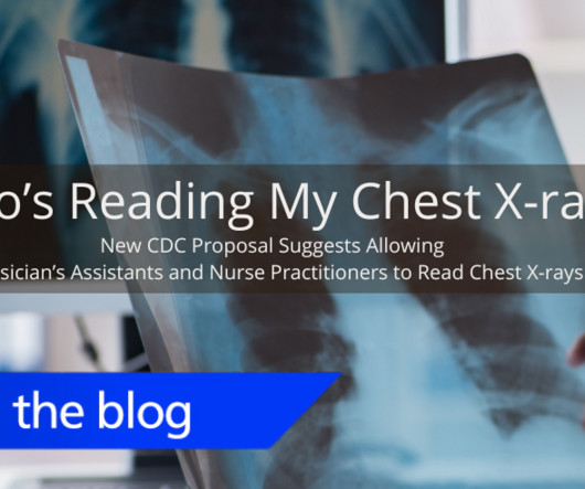
This program trains and certifies physicians to read chest radiographs for workers participating in health surveillance programs. Radiologists complete over 20,000 hours of clinical experience before being certified to interpret radiographic images. Compared to only 500 hours of clinical experience for NPs and 2000 hours for PAs.
Representative chest radiographs and graphs of lung signal intensity obtained using DDR for the severe COPD group. a) Radiograph during forced inspiration when the average signal intensity reached its maximum (SI max = 3879.3). (b) The subject is a 77-year-old male with VC of 1.68 L and FEV1 of 0.68
He earned his medical degree at the University of Alabama School of Medicine and completed a residency in diagnostic radiology at the University of Florida. An assistant professor of radiology and radiological science, Francis Deng, MD, is the Johns Hopkins radiology department's co-director of medical student diagnostic radiology electives.
He earned his medical degree at the University of Alabama School of Medicine and completed a residency in diagnostic radiology at the University of Florida. I'm a radiographer,' " Stewart recalled. I thought, 'I'm not a teacher. But Hennessy encouraged her.
milla1cf Fri, 07/07/2023 - 21:32 July 7, 2023 — FUJIFILM Healthcare Americas Corporation, a leading provider of diagnostic and enterprise imaging solutions, today announced the U.S. X-rays are the most widely used diagnostic tests, accounting for 60% of all imaging studies conducted.
Substituting less energy-consuming ultrasound for x-ray or CT reduced energy use by as much as 8% during diagnostic radiology processes and 31.2% for total CFCs, while demonstrating an increase in diagnostic activity of 11.8%, Masperi said. Stage two consisted of a retrospective review of exams performed and their timing.
Understanding the mechanics of flow artifacts on CT or CT angiography (CTA) and how these artifacts are created is key to better disease diagnosis, according to a review published April 25 in RadioGraphics. Systemic arterial flow artifacts can cause diagnostic confusion with dissections, vascular injuries, and thrombi.
The group's commentary was published February 13 in RadioGraphics. Although midfield MRI systems show promise for diagnostic imaging, they will require more software and hardware development, wrote Vivek Pai, MD, and Pejman Jabehdar Maralani, MD, both of University of Toronto in Canada, in an accompanying commentary.
Shamie Kumar describes how AI fits into a radiology clinical workflow and her perspective on how a clinical radiographer could use this to learn from and enhance their skills. If the AI findings are seen in PACS, how many radiographers actually log into PACS after taking a scan or X-ray? Can Radiographers Up-Skill?
This competition demonstrated the value of AI in detecting and localizing many pathologies in chest radiographs by simulating the real work situations of radiologists,” the group wrote. The performance of all radiologists with and without AI assistance showed that AI improved the diagnostic accuracy of the doctors,” the group wrote.
In an open forum, Yi Xiang Tay, of Singapore University Hospital's radiography and diagnostic imaging department, shared his team's research. It was the endnote of a series of sessions focused on optimizing radiology services.
To bridge this knowledge gap, the group analyzed 327 PET/CT diagnostic radiology reports between January 2001 and December 2018 at their institution that included descriptions of the finding. These signs often prompt otolaryngology referral to rule out malignancy yet the true risk based on the finding is unknown, they noted.
Healthcare disparities continue to plague medical imaging, but there are concrete measures radiologists can take to mitigate them, according to a paper published on October 12 in RadioGraphics. Image courtesy of Peter Abraham, MD, et al.
Common diagnostic tests for pulmonary disorders include chest x-rays and pulmonary function tests (PFTs). a) Raw example of a dynamic digital radiograph. (b) The study was published March 29 in Chest Pulmonary. Although essential, these tests offer a limited static assessment, the authors wrote.
This ensures an unobstructed qualitative evaluation, and enables a quantitative evaluation of dark-field radiographs,” Urban et al wrote. Dark-field chest x-ray has just recently been translated to the clinical stage and its diagnostic value is currently being investigated with a clinical prototype, the authors wrote.
The study, published in the journal Diagnostics, highlights the clinical value of DDR through its unique ability to evaluate diaphragm movement in real time and integrate dynamic functional information with static anatomical data to provide a quantitative assessment of diaphragmatic movement, including excursion and speed.
milla1cf Wed, 06/12/2024 - 21:59 June 12, 2024 — Carestream launched its Image Suite MR 10 Software to help deliver a boost to productivity and efficiency while enabling a more user-friendly imaging experience for radiographers. Image Suite MR 10 helps push the envelope further for even more focus on patient care.”
"Together with our radiographers, I learned to scan cardiac patients and learned special anatomy from pediatric cardiologists and pediatric cardiac surgeons." He noted that in 2019, the European Society of Cardiology issued updated guidelines for diagnostic imaging of coronary artery disease (CAD), recommending noninvasive imaging (i.e.,
Whenever bilateral standing radiographs would have been needed, a WBCT was performed instead. This was the case for diagnostic purposes but also for follow-up imaging at 3, 6 and 12 months after re-alignment procedures, fusions or arthroplasties. I use WBCT every day.
11, 2025 Harrison.ai, a developer of AI-powered medical diagnostic support and workflow solutions, has announced the accelerated expansion of its operations into the United States a move supported by US$112 million of Series C funding. Radiologists using Harrison.ai's technology have seen an over 45% increase in diagnostic accuracy1.
Decades since the advent of breast scanning technology, innovations in noninvasive diagnostic imaging provide new options in the field of early detection. However, overcompression artificially lowers the radiographic density. A mammogram can show how dense breasts are including how low or high in density.
Teleradiology-India Introduction: “X-ray Visionaries” takes you on a compelling journey to unveil the expertise of radiographers and technologists, the unsung heroes of X-ray technology. Chapter 1: Introduction to Radiographers and Technologists An overview of the pivotal roles radiographers and technologists play in healthcare.
Radiology plays a crucial role in modern medicine, answering essential diagnostic questions pertaining to nearly every pathology facing clinicians. At seven days post-imaging, nearly half had unreported brain and chest CT scans, while 59% had unreported chest radiographs. The consequences are significant.
A 2023 study for example found that ChatGPT-4 performed well on the image-independent American College of Radiology (ACR) Diagnostic In-Training Exam (ACR DXIT) practice questions. ChatGPT has shown mixed results when taking different exams from various radiologic societies.
Our hope is that we'll be able to determine how to use this as a diagnostic tool in actual practice,” he said. He added that by the age of 50 or 60, many or most people have already had a CT scan for diagnostic purposes. “We We can first take advantage of all those scans,” he said.
Topics include the following: Lunit's AI model combines new and existing AI algorithms to filter normal chest radiographs from the radiology workload An exploration of the accuracy and robustness of Lunit's Insight MMG compared with radiologist readers Lunit's AI model tracks mammographic parenchymal patterns as predictive markers for breast cancer (..)
The department of radiology teaches a diagnostic technologies in healthcare course, principally around imaging technologies, for which 3,000 people have signed up, Rubin said. "It
In pediatric imaging, anesthesia is commonly thought to be necessary for obtaining high-quality diagnostic imaging, but an innovative program at the UCSF is challenging that idea.
When reviewing radiographs, computed tomography (CT) scans or magnetic resonance imaging (MRI) scans, do you still turn to mnemonics every now and then to jog your short-term memory?
The authors note that ionizing radiation is the basis for the production of diagnostic X-rays, however it has long been proven to increase the risk of cancer. 26 –29, 2023 in North Hall, Level 3, booth #7913.
Repeating imaging exams increases the workload of your radiographers who are already stretched too thin; increases the exposure of the affected patients; and contributes to patients’ reduced confidence and satisfaction with your imaging department. The Audio Assist makes it easier for radiographers to hear the patients.
In medical imaging and diagnostics, advanced image recognition has profound applications in medical imaging and diagnostics. AI tools that enable radiographers to separate noise from an image already exist today. Carestream’s Smart Assist features can help you ease the burden on your radiographers.
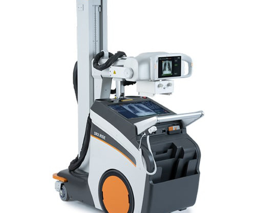
Additionally, Carestream will display: Carestream’s full line of DR detectors — state-of-the-art DR detector options that are available in a variety of sizes to fit a facility’s workflow and budget, including the award-winning, glass-free Lux 35 Detector and small-format DRX Plus 2530C Detector.
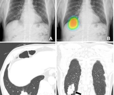
A CT-based radiomic model offered over 10 percent higher specificity and positive predictive value for high-risk lung adenocarcinoma in comparison to a radiographic model, according to external validation testing in a recent study.
The Society publishes five established journals: Radiology, RadioGraphics, Radiology: Artificial Intelligence, Radiology: Cardiothoracic Imaging and Radiology: Imaging Cancer. RadioGraphics , RSNA’s premier educational journal, edited by Christine ‘Cooky’ Menias , M.D., Klein , M.D., RSNA Board Liaison for Publications.
MyImageDx facilitates the delivery of learning world-wide for peer review, MDT and discrepancy activities for students, radiologists and radiographers anywhere, anytime. MyImageDx providers radiology educators access to a personalized imaging educational platform without having to build it in-house.
We organize all of the trending information in your field so you don't have to. Join 5,000 users and stay up to date on the latest articles your peers are reading.
You know about us, now we want to get to know you!
Let's personalize your content
Let's get even more personalized
We recognize your account from another site in our network, please click 'Send Email' below to continue with verifying your account and setting a password.


Let's personalize your content