This site uses cookies to improve your experience. To help us insure we adhere to various privacy regulations, please select your country/region of residence. If you do not select a country, we will assume you are from the United States. Select your Cookie Settings or view our Privacy Policy and Terms of Use.
Cookie Settings
Cookies and similar technologies are used on this website for proper function of the website, for tracking performance analytics and for marketing purposes. We and some of our third-party providers may use cookie data for various purposes. Please review the cookie settings below and choose your preference.
Used for the proper function of the website
Used for monitoring website traffic and interactions
Cookie Settings
Cookies and similar technologies are used on this website for proper function of the website, for tracking performance analytics and for marketing purposes. We and some of our third-party providers may use cookie data for various purposes. Please review the cookie settings below and choose your preference.
Strictly Necessary: Used for the proper function of the website
Performance/Analytics: Used for monitoring website traffic and interactions
has developed neural networks for interpreting chest x-rays that could help speed the adoption of AI systems in clinical settings, according to a study published December 8 in The Lancet Digital Health. To that end, the group first compiled the largest chest x-ray data set to date in the U.K. million frontal x-rays.
A team in Europe has launched a novel web application to help radiology trainees gain skills in detecting lung nodules on chest x-rays. To that end, Borgbjerg and colleagues aimed to develop and make available a pure web-based application for perception training in lung nodule detection in chest x-rays.
In addition, the company discussed expansion of its SilverBeam x-ray beam shaping energy filter. After clinicians are informed of these urgent situations, Canons Mobile DICOM Viewer offers remote access to images and results. Food and Drug Administration 510(k) clearance).
These sessions, which will be held in the Lakeside Learning Center, will cover a wide range of AI topics, such as basics of natural language processing in radiology, data processing and curation for deep learning, and using ChatGPT for DICOM deidentification. 10:10 a.m. | 10:10 a.m. | 9:00 a.m. |
The Belgium-based company Osimis was created following the success of Orthanc, an open-source DICOM server for healthcare and medical research, according to its website. Osimis has since become a vendor-neutral AI integration platform for radiology departments.
DICOM Image Format is an international standard to transmit, store, retrieve, print, process, and display medical imaging information. DICOM allows transmitting medical imaging data to devices like scanners, servers, workstations, printers, network hardware, and PACS. What is DICOM Image Format?
Introduction: The marriage of cloud technology with Picture Archiving and Communication Systems (PACS) and Digital Imaging and Communications in Medicine (DICOM) standards has ushered in a new era for medical imaging services. Explore the flexibility in managing data from various sources, including X-rays, MRIs, CT scans, and more.
German experts shared their experience with the “AI to SR pipeline,” which integrates a commercially available AI tool for chest X-ray pathology detection and localization into structured report templates.
X-ray is the oldest form of imaging and a valuable tool on the frontlines of patient care, accounting for over 60 percent of all imaging exams. [4] X-ray is the oldest form of imaging and a valuable tool on the frontlines of patient care, accounting for over 60 percent of all imaging exams. [4]
Download our: Medical Imaging in Oncology Review brochure here DICOM, PACS, RIS, EHRs, HIS, VNAs Accessibility and efficiency have been key drivers of innovation (as well as a multitude of acronyms!) VNAs are bound by a general agreement on how DICOM images are stored and accessed via a networked interface. CT scans, X-rays).
Diagnosis often depends on the interpretation of tests like of X-Rays, CT Scan, MRI, PET CT Scan , etc. Digital Imaging and Communications in Medicine (DICOM) is used to transmit and store data. It has adhered to the globally standardized reporting format of X-Rays, CT Scan, MRI, PET CT Scan, etc.
The teleradiology services provider delivers accurate timely radiology interpretation of the X-ray exams they receive from the 500+ Concentra locations nationwide. This approach allows Premier to work with providers like Concentra with highly customized workflows through DICOM scripting and HL7 integration.”
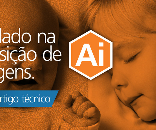
Soluções pediátricas para aquisição de imagens Receptor de imagem: Capturar a imagem de raios X com o receptor de imagem é a primeira etapa da formação da imagem. Software SmartGrid: A dispersão de raios X poderá comprometer consideravelmente a qualidade da imagem se não for gerenciada como parte do processo de aquisição.
La introducción de los productos detectores DRX inalámbricos de Carestream ha sido un gran paso adelante en la provisión de un detector de rayos X de alta calidad que se adapta perfectamente al flujo de trabajo de la UCIN y la UCI pediátrica. link] (Consultado el 2012-09-27). link] (Consultado el 12 de julio de 2022).
We organize all of the trending information in your field so you don't have to. Join 5,000 users and stay up to date on the latest articles your peers are reading.
You know about us, now we want to get to know you!
Let's personalize your content
Let's get even more personalized
We recognize your account from another site in our network, please click 'Send Email' below to continue with verifying your account and setting a password.


Let's personalize your content