This site uses cookies to improve your experience. To help us insure we adhere to various privacy regulations, please select your country/region of residence. If you do not select a country, we will assume you are from the United States. Select your Cookie Settings or view our Privacy Policy and Terms of Use.
Cookie Settings
Cookies and similar technologies are used on this website for proper function of the website, for tracking performance analytics and for marketing purposes. We and some of our third-party providers may use cookie data for various purposes. Please review the cookie settings below and choose your preference.
Used for the proper function of the website
Used for monitoring website traffic and interactions
Cookie Settings
Cookies and similar technologies are used on this website for proper function of the website, for tracking performance analytics and for marketing purposes. We and some of our third-party providers may use cookie data for various purposes. Please review the cookie settings below and choose your preference.
Strictly Necessary: Used for the proper function of the website
Performance/Analytics: Used for monitoring website traffic and interactions
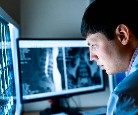
Introduction: The marriage of cloud technology with PictureArchiving and CommunicationSystems (PACS) and DigitalImaging and Communications in Medicine (DICOM) standards has ushered in a new era for medical imaging services.
All this is possible because of PACS. PACS – PictureArchiving and CommunicationSystem; a system involved in acquiring the medical images, transmission, viewing, storage, and retrieval of same images. Since 1980, PACS has undergone many transformations in multiple ways.
A Vendor Neutral Archive (VNA) is a medical imaging technology that provides a centralized storage solution for medical images and associated data, regardless of the specific imaging devices or systems used to generate those images. What is Vendor Neutral Archive (VNA)? What is PACS?
Introduction: “Radiology Reshaped: The Digital Transformation Unveiled” takes you on a journey into the profound changes occurring in the field of radiology as it undergoes a comprehensive digital transformation. Highlight key milestones that paved the way for the digital transformation.
Medical images management: how implementing proper processes and quality control can ensure maximum operational effectiveness and regulatory compliance Between February 2018 and March 2019, ~44.9 million diagnostic imaging tests were performed in England [i]. in managing images, and several components have evolved.
tim.hodson Fri, 08/09/2024 - 15:45 At the annual AHRA (American Healthcare Radiology Administrators) conference in Orlando, Florida, Bayer announced an exploratory collaboration with Alara Imaging, Inc. Alara is proud to collaborate with Bayer to support health systems across the U.S. JAMA Internal Medicine. Jan 2019;364:k4931.
Teleradiology services in India stand as a shining example, harnessing innovation in imaging technology to revolutionize the way diagnostic information is acquired, interpreted, and shared. **1. DigitalImaging Revolution: From Film to Pixels: Explore the shift from traditional film-based imaging to digital radiography.
Teleradiology Introduction: In the landscape of modern medicine, teleradiology stands as a technological powerhouse, revolutionizing the way medical images are acquired, interpreted, and shared. DigitalImaging Modalities: Discuss the transition from traditional film to digitalimaging modalities.
Introduction : Teleradiology is a branch of telemedicine in which telecommunications systems are used to transmit radiological images and related data from one location to another for diagnostic and consulting purposes. Moreover, the images can be accessed from different places hassle-free. from 2018 to 2025.
In the ever-evolving landscape of modern medicine, technology plays a crucial role in transforming patient care, enhancing diagnostic accuracy, and streamlining workflows. CPACS stands for Cardiovascular PictureArchive and CommunicationSystem. One such pivotal technology in cardiology is CPACS.
After working for weeks in his lab experimenting on the production of ‘strange rays’, which he referred to as ‘X’, he asked his wife Anna Bertha to lend ‘a hand’, the left one to be precise, which he used to produce the first X-ray image. after seeing the image. (2) It all started when Wilhelm Conrad Röntgen discovered X-rays in 1895.
We organize all of the trending information in your field so you don't have to. Join 5,000 users and stay up to date on the latest articles your peers are reading.
You know about us, now we want to get to know you!
Let's personalize your content
Let's get even more personalized
We recognize your account from another site in our network, please click 'Send Email' below to continue with verifying your account and setting a password.


Let's personalize your content