This site uses cookies to improve your experience. To help us insure we adhere to various privacy regulations, please select your country/region of residence. If you do not select a country, we will assume you are from the United States. Select your Cookie Settings or view our Privacy Policy and Terms of Use.
Cookie Settings
Cookies and similar technologies are used on this website for proper function of the website, for tracking performance analytics and for marketing purposes. We and some of our third-party providers may use cookie data for various purposes. Please review the cookie settings below and choose your preference.
Used for the proper function of the website
Used for monitoring website traffic and interactions
Cookie Settings
Cookies and similar technologies are used on this website for proper function of the website, for tracking performance analytics and for marketing purposes. We and some of our third-party providers may use cookie data for various purposes. Please review the cookie settings below and choose your preference.
Strictly Necessary: Used for the proper function of the website
Performance/Analytics: Used for monitoring website traffic and interactions
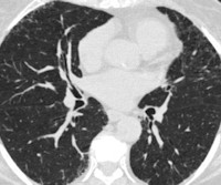
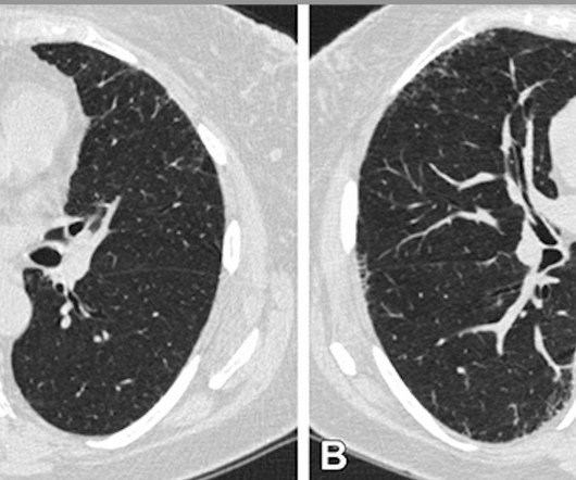
CT imaging shows that severe acute respiratory disease events can be caused by quantitative interstitial abnormalities (QIA) -- that is, small irregularities that don't necessarily meet diagnostic criteria for advanced pulmonary diseases but show up on CT exams over time, a study published April 30 in Radiology has reported.
Millions of CT scans are performed each year -- often for lung cancer screening, but also as a first-line test when a patient presents in the emergencyroom -- and information from these tests can shed light on other conditions. AI can perform coronary artery disease risk stratification Sunday, December 1 | 3:20 p.m.-3:30
Appropriate candidates for this procedure are patients with a low- to intermediate- probability of significant coronary artery disease (for example, a 45- to 55-year-old patient who presents with atypical chest pain). If the CCTA exam is negative, the patient can be sent home and treated as an outpatient.
Emergencyrooms (ERs) play a critical and indispensable role in the healthcare system, serving as the front line of medical care for individuals experiencing urgent and life-threatening situations. During the summer months in the United States, emergencyrooms tend to see an increase in patient visits due to various reasons.
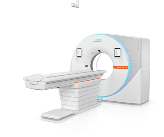
The Naeotom Alpha.Prime is the world’s first single source photon-counting CT for use as a high-performance scanner for in-patient, ambulatory, and emergencyroom examinations in stand-alone institutions and big IDNs (Integrated Delivery Networks), or also in the periphery of hub-and-spoke networks.
DDR could also provide a more comprehensive understanding of a patient’s neuromuscular component of ventilatory disorders and many other pulmonary diseases.” We are excited about the ability to deploy this in emergencyroom, inpatient and intensive care medicine.
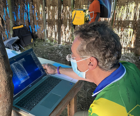
Rob Liddell, MD, is a diagnostic radiologist who used MinXray’s Impact Wireless system to take radiographic images of patients in the village and screen for common diseases in the region, such as tuberculosis and emphysema. He also used the system to diagnose cancers, infections and various musculoskeletal injuries.
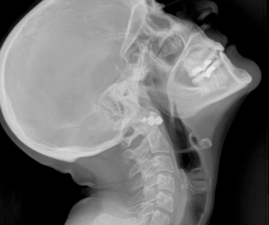
Researchers have found that nonchest CT exams taken in the emergencyroom for other indications may also capture pulmonary findings suspicious for COVID-19, according to a study published May 11 in Radiology. Of the 119 patients, 101 had abdomen/pelvis CT, and 18 had cervical spine/neck CT. Images courtesy of the RSNA.
Several factors contribute to delays and overcrowding in the emergency department, such as: The increasing demand for emergency services, driven by factors such as population growth, aging demographics and limited access to primary care services.
Trauma patients are common, some coming directly from the emergencyroom. Locum Tenens Radiology with Mint Physician Staffing There are many factors that are increasing the demand for radiology, including the aging population and the growth of chronic diseases and cancer survivors. 3 Who Is Treated in Radiology?
CT scans are faster than MRIs and can be crucial in emergencies. Uses of CT Scans: Emergency Diagnoses: Quick to perform, they are used in emergencyrooms to diagnose internal injuries and bleeding. Dental Assessment: Helps in diagnosing tooth decay and periodontal disease.
However, evaluating and managing patients with acute alcohol intoxication in the emergency department can be challenging. The situation is more dangerous given the high incidence of chronic disease, critical illness, and acute trauma. Annals of Emergency Medicine. Alcoholic Liver Disease: Pathogenesis and Current Management.
milla1cf Fri, 05/24/2024 - 07:00 May 24, 2024 — Smokers who have small abnormalities on their CT scans that grow over time have a greater likelihood of experiencing acute respiratory disease events, according to a new study published today in Radiology , a journal of the Radiological Society of North America ( RSNA ).
Our heightened emphasis on the North American marketplace solidifies our commitment to making a meaningful impact in the fight against this deadly disease and underscores our dedication to advancing healthcare through innovation, providing a transformative solution enhancing the early detection of cancer and ultimately improving patient outcomes.”
Alzheimer’s disease is the most common and well known rendition of dementia, but there are other forms of the disease as well. Small Vessel Disease / Vascular Dementia The brain is full of small arteries that supply necessary oxygen-rich blood and nutrients.
Recent evidence supports that patients experiencing vascular emergencies like stroke make up some of the most dangerous errors, contributing up to 17.5% of the serious harm rate calculated among dangerous disease cases. Likewise, severity is often underestimated due to variable and non-specific symptomology and localization.
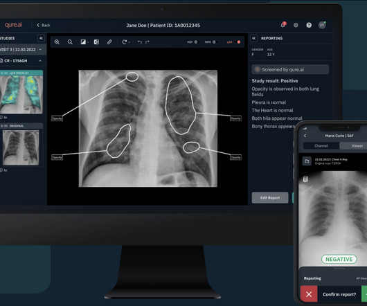
The findings boost AI-driven medical diagnostics and bring health care professionals closer to being able to quickly diagnose patients with COVID-19 and other pulmonary diseases with algorithms that comb through ultrasound images to identify signs of disease.
to be exact) that the patient actually has the disease. to be exact) that the patient actually has the disease. But tennis is still very much on the kind end of the spectrum compared to, say, a hospital emergencyroom, where doctors and nurses do not automatically find out what happens to a patient after their encounter.
We organize all of the trending information in your field so you don't have to. Join 5,000 users and stay up to date on the latest articles your peers are reading.
You know about us, now we want to get to know you!
Let's personalize your content
Let's get even more personalized
We recognize your account from another site in our network, please click 'Send Email' below to continue with verifying your account and setting a password.


Let's personalize your content