This site uses cookies to improve your experience. To help us insure we adhere to various privacy regulations, please select your country/region of residence. If you do not select a country, we will assume you are from the United States. Select your Cookie Settings or view our Privacy Policy and Terms of Use.
Cookie Settings
Cookies and similar technologies are used on this website for proper function of the website, for tracking performance analytics and for marketing purposes. We and some of our third-party providers may use cookie data for various purposes. Please review the cookie settings below and choose your preference.
Used for the proper function of the website
Used for monitoring website traffic and interactions
Cookie Settings
Cookies and similar technologies are used on this website for proper function of the website, for tracking performance analytics and for marketing purposes. We and some of our third-party providers may use cookie data for various purposes. Please review the cookie settings below and choose your preference.
Strictly Necessary: Used for the proper function of the website
Performance/Analytics: Used for monitoring website traffic and interactions
To cover a CT and MR "list" was a luxury; it was to escape the murderous fluoroscopy rooms, intravenous urography (IVU) lists, and indeterminable piles of plain films Then the scanners got fast. So as to report more CT and MRI, radiologists stopped doing hands-on ultrasound and fluoroscopy. 20s rotation, 40s reconstruction.
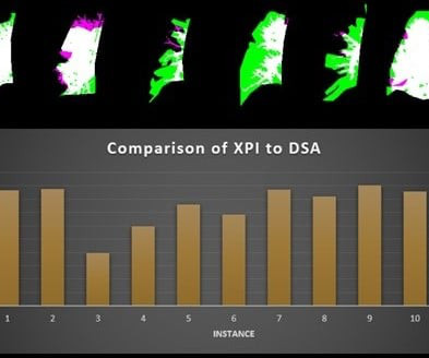
A fluoroscopy method incorporating a noncontrast x-ray pulsatility index (XPI) can improve clinical efficiency as a screening or diagnostic test for suspected chronic thromboembolic pulmonary hypertension, according to research presented at the American Roentgen Ray Society (ARRS) annual meeting.
. | M7-SSPH05-2 | Room N229 Findings will be presented in this Monday afternoon presentation on organ-specific ionizing radiation doses in neonatal patients who undergo interventional procedures for congenital heart disease (CHD). Louis, and colleagues.
Imaging more broadly offers significant value to patients, providers, and health care systems when used appropriately through disease prevention, detection, diagnosis, prognosis, and in the delivery and monitoring of precise, minimally invasive treatment," the group explained. X-ray or fluoroscopy 0.82 Nuclear medicine 0.76
echocardiography) for congenital heart disease, diagnostic; 76987 including placement and manipulation of transducer, image acquisition, interpretation, and report 76988 placement, manipulation of transducer, and image acquisition only. epiaortic), diagnostic Intraoperative epicardial cardiac ultrasound (i.e.,
Ultrasound model predicts liver disease progression. Commercially Available Chest Radiograph AI Tools for Detecting Airspace Disease, Pneumothorax, and Pleural Effusion. Increased Metabolic Activity of the Thymus and Lymph Nodes in Pediatric Oncology Patients After Coronavirus Disease 2019 Vaccination.
The new imaging systems that will be on display include three new digital radiography (DR) suites, two new fluoroscopy systems, a 0.4T Persona C-HR: Fujifilm’s newest mobile fluoroscopy c-arm solution providing 30 frames per second FPS pulsed fluoroscopy images at low dose. The sturdy patient table features a robust 800 lbs.
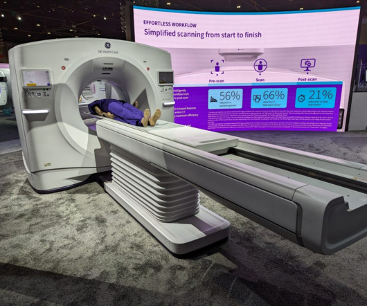
The research will produce technical feedback to assist GEHC in assessing the system’s reconstruction methods, image presentation workflow, and clinical benefits for specific pathologies and disease types. MRI GEHC unveiled Signa Champion, a 1.5-tesla,
Ultimately, in this ARRS Annual Meeting Cum Laude Scientific Poster , where noncontrast (XPI) and contrast pulmonary angiography images were obtained in 11 different lungs, all patients were able to perform satisfactory breath hold, despite moderate to severe disease.
She believes that when you begin interacting with patients during fluoroscopy or interventional procedures, it fosters the growth of empathy. We should be leveraging the ‘free’ body composition data that is available on all abdominal CTs.”— This empathy, she contends, significantly influences the way you generate reports.
Unlike fluoroscopy, DDR is a series of individual digital X-ray images acquired at high speed and low doses, allowing the visualization of anatomy in motion. In addition to the lower dose that X-ray delivers compared to fluoroscopy, patients also benefit from the increased distance between them and the X-ray detector.
Radiology is a medical imaging procedure that uses ionizing electromagnetic radiation to create images of bones, organs, and soft tissues to diagnose a patient’s symptoms, disease, or conditions. It includes techniques like X-rays, CT scans, MRIs, ultrasounds, and fluoroscopy.
CT LVAS, designed for use with computed tomography scans, joins 4D Medical's XV LVAS imaging software cleared for use with fluoroscopy in the United States. Standard CT images provide clinicians with structural detail about patients' lungs—from which they try to infer information about disease and function.
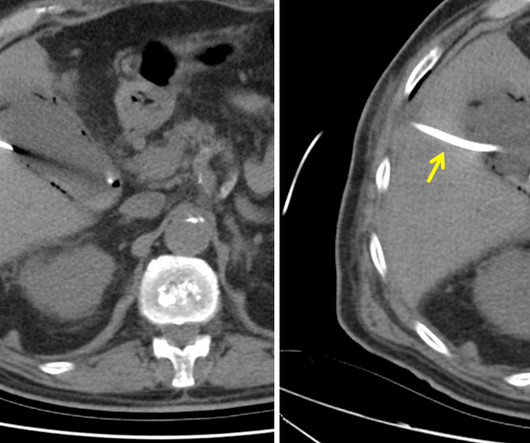
Primary hepatic gas gangrene is a rare but potentially fatal infectious disease (1). This letter reports a primary hepatic gas gangrene case successfully treated with fluoroscopy-guided percutaneous drainage. The role of interventional procedures in managing hepatic gas gangrene is limited in the literature.
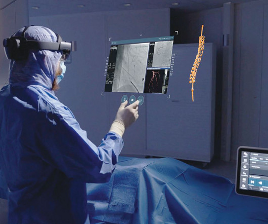
Image-guided therapy is a fast-evolving space that leverages advanced imaging and visualization solutions to make it possible to treat many conditions, from cancer care to cardiovascular disease and more, with little to no surgical incisions.
Fluoroscopy: Real-Time Insights into Heart Function: Fluoroscopy is another dynamic application of X-ray technology, allowing healthcare professionals to monitor the real-time function of the heart and lungs. It’s an essential tool for detecting coronary artery disease and pulmonary embolisms.
Introduction: Radiology is a gateway to the invisible, offering a window into the human body and enabling a deeper understanding of health and disease. Radiological Milestones: Discuss key milestones in the history of radiology, from the advent of fluoroscopy to the development of advanced imaging techniques.
Chapter 3: The Radiologic Toolbox – Types of X-ray Imaging An exploration of the various types of X-ray imaging, including radiography, fluoroscopy, computed tomography (CT), and more. How X-rays are generated, interact with human tissue, and create diagnostic images. How each modality serves unique clinical purposes and applications.
Chapter 3: Types of X-ray Imaging: Beyond Radiography An exploration of the various types of X-ray imaging, including radiography, fluoroscopy, and computed tomography (CT). The principles of radiation and how X-rays interact with the human body to create diagnostic images. How each modality is used for different clinical purposes.
Rarer causes include rheumatic disease, congenital, myxomatous degeneration, endocarditis, or pulmonary hypertension [3]. The procedure first begins with gaining femoral/jugular vein access and inserting a stiff guidewire into the right atrium, confirmed with fluoroscopy [5,6]. The leading cause of TR is due to left heart failure.
The patient is pregnant (orange arrows), therefore ionizing radiation with CT scan or fluoroscopy can not be used for imaging guidance. Interventional approaches to gallbladder disease. A transperitoneal route is favored in patients with coagulopathy or diffuse liver disease [1]. MRI of abdomen. Case Rep Gastroenterol.
We organize all of the trending information in your field so you don't have to. Join 5,000 users and stay up to date on the latest articles your peers are reading.
You know about us, now we want to get to know you!
Let's personalize your content
Let's get even more personalized
We recognize your account from another site in our network, please click 'Send Email' below to continue with verifying your account and setting a password.


Let's personalize your content