This site uses cookies to improve your experience. To help us insure we adhere to various privacy regulations, please select your country/region of residence. If you do not select a country, we will assume you are from the United States. Select your Cookie Settings or view our Privacy Policy and Terms of Use.
Cookie Settings
Cookies and similar technologies are used on this website for proper function of the website, for tracking performance analytics and for marketing purposes. We and some of our third-party providers may use cookie data for various purposes. Please review the cookie settings below and choose your preference.
Used for the proper function of the website
Used for monitoring website traffic and interactions
Cookie Settings
Cookies and similar technologies are used on this website for proper function of the website, for tracking performance analytics and for marketing purposes. We and some of our third-party providers may use cookie data for various purposes. Please review the cookie settings below and choose your preference.
Strictly Necessary: Used for the proper function of the website
Performance/Analytics: Used for monitoring website traffic and interactions
To cover a CT and MR "list" was a luxury; it was to escape the murderous fluoroscopy rooms, intravenous urography (IVU) lists, and indeterminable piles of plain films Then the scanners got fast. So as to report more CT and MRI, radiologists stopped doing hands-on ultrasound and fluoroscopy. 20s rotation, 40s reconstruction.
Cardiac Intraoperative Ultrasound (IOUS) Services : New codes are available to report cardiac IOUS, as follows: CPT Code Description 76984 Ultrasound, intraoperative thoracic aorta (e.g., epiaortic), diagnostic Intraoperative epicardial cardiac ultrasound (i.e., Ultrasound guidance is now bundled with the primary procedure.
Higher Medicaid-to-Medicare reimbursement ratios (MMRR) are linked to increased likelihood of Medicaid patients receiving CT, MR, ultrasound, and x-ray imaging, researchers have reported. Ultrasound 0.85 X-ray or fluoroscopy 0.82 higher for ultrasound, and 31.8% Nuclear medicine 0.76 higher for MR, 21.4% higher for x-ray.
Ultrasound model predicts liver disease progression. Appropriateness and imaging outcomes of ultrasound, CT, and MR in the emergency department: a retrospective analysis from an urban academic center. Commercially Available Chest Radiograph AI Tools for Detecting Airspace Disease, Pneumothorax, and Pleural Effusion.
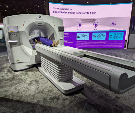
The research will produce technical feedback to assist GEHC in assessing the system’s reconstruction methods, image presentation workflow, and clinical benefits for specific pathologies and disease types. Other ultrasound scanners on display included GEHC’s Logiq E10, Fortis, Invenia ABUS 2.0, MRI GEHC unveiled Signa Champion, a 1.5-tesla,
The new imaging systems that will be on display include three new digital radiography (DR) suites, two new fluoroscopy systems, a 0.4T Persona C-HR: Fujifilm’s newest mobile fluoroscopy c-arm solution providing 30 frames per second FPS pulsed fluoroscopy images at low dose. The sturdy patient table features a robust 800 lbs.
Radiology is a medical imaging procedure that uses ionizing electromagnetic radiation to create images of bones, organs, and soft tissues to diagnose a patient’s symptoms, disease, or conditions. It includes techniques like X-rays, CT scans, MRIs, ultrasounds, and fluoroscopy.
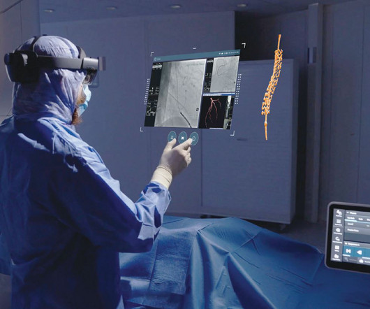
Image-guided therapy is a fast-evolving space that leverages advanced imaging and visualization solutions to make it possible to treat many conditions, from cancer care to cardiovascular disease and more, with little to no surgical incisions.
Introduction: Radiology is a gateway to the invisible, offering a window into the human body and enabling a deeper understanding of health and disease. Radiological Milestones: Discuss key milestones in the history of radiology, from the advent of fluoroscopy to the development of advanced imaging techniques.
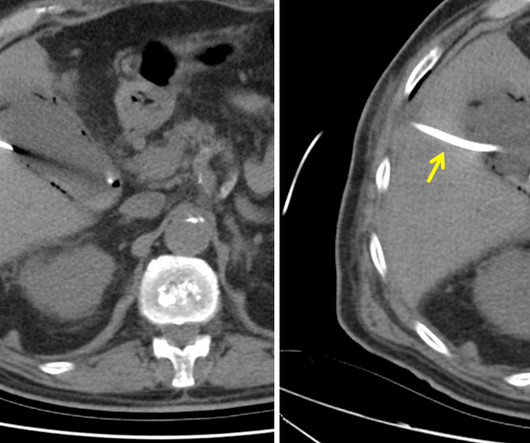
The patient is pregnant (orange arrows), therefore ionizing radiation with CT scan or fluoroscopy can not be used for imaging guidance. Ultrasound of gallbladder used for guidance of percutaneous needle (red arrow) placement for cholecystostomy. Ultrasound of gallbladder demonstrating drainage catheter in the lumen (blue arrow).
We organize all of the trending information in your field so you don't have to. Join 5,000 users and stay up to date on the latest articles your peers are reading.
You know about us, now we want to get to know you!
Let's personalize your content
Let's get even more personalized
We recognize your account from another site in our network, please click 'Send Email' below to continue with verifying your account and setting a password.


Let's personalize your content