This site uses cookies to improve your experience. To help us insure we adhere to various privacy regulations, please select your country/region of residence. If you do not select a country, we will assume you are from the United States. Select your Cookie Settings or view our Privacy Policy and Terms of Use.
Cookie Settings
Cookies and similar technologies are used on this website for proper function of the website, for tracking performance analytics and for marketing purposes. We and some of our third-party providers may use cookie data for various purposes. Please review the cookie settings below and choose your preference.
Used for the proper function of the website
Used for monitoring website traffic and interactions
Cookie Settings
Cookies and similar technologies are used on this website for proper function of the website, for tracking performance analytics and for marketing purposes. We and some of our third-party providers may use cookie data for various purposes. Please review the cookie settings below and choose your preference.
Strictly Necessary: Used for the proper function of the website
Performance/Analytics: Used for monitoring website traffic and interactions
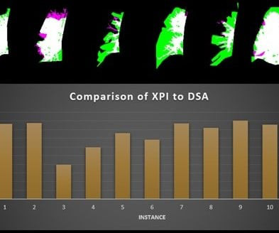
A fluoroscopy method incorporating a noncontrast x-ray pulsatility index (XPI) can improve clinical efficiency as a screening or diagnostic test for suspected chronic thromboembolic pulmonary hypertension, according to research presented at the American Roentgen Ray Society (ARRS) annual meeting.
Higher Medicaid-to-Medicare reimbursement ratios (MMRR) are linked to increased likelihood of Medicaid patients receiving CT, MR, ultrasound, and x-ray imaging, researchers have reported. X-ray or fluoroscopy 0.82 higher for x-ray. Nuclear medicine 0.76 Ultrasound 0.85 higher for MR, 21.4%
. | M7-SSPH05-2 | Room N229 Findings will be presented in this Monday afternoon presentation on organ-specific ionizing radiation doses in neonatal patients who undergo interventional procedures for congenital heart disease (CHD). Louis, and colleagues.
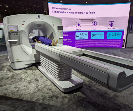
The scanner also comes with True Enhance DL, an AI-based application that generates deep learning-based monochromatic-like images from a single-energy x-ray acquisition. Another introduction, Definium Pace, is a fixed x-ray system available at a value price point, according to GEHC.
Introduction: Radiology is a gateway to the invisible, offering a window into the human body and enabling a deeper understanding of health and disease. “X-Rays and Beyond” embarks on an exciting journey to explore the captivating world of radiology, highlighting how it goes far beyond just X-rays.
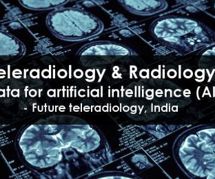
Teleradiology & Radiology data for artificial intelligence (AI) Introduction: “Illuminating Shadows” invites you on a comprehensive journey into the fascinating world of X-ray imaging. Chapter 1: Introduction to X-ray Imaging An overview of the importance of X-ray imaging in healthcare.
Closeup of X-ray photography of human brain Introduction: “The Radiant Thread” is a comprehensive guide that unravels the intricate world of X-ray imaging from A to Z. From its historical roots to contemporary innovations, we will follow the radiant thread that connects all aspects of X-ray imaging.
announced significant expansion of X-Ray systems with Dynamic Digital Radiography (DDR) at multiple healthcare institutions across the US, including at Appleton Area Health (Appleton, Minn.), A powerful X-ray generator and Automatic Exposure Control further optimize image quality and help minimize patient dose.
The new imaging systems that will be on display include three new digital radiography (DR) suites, two new fluoroscopy systems, a 0.4T Persona C-HR: Fujifilm’s newest mobile fluoroscopy c-arm solution providing 30 frames per second FPS pulsed fluoroscopy images at low dose. The sturdy patient table features a robust 800 lbs.
Additionally,” said Matthew Smith , MD, from Vanderbilt University Medical Center in Nashville, TN, “this easy-to-implement method can be performed by an x-ray technologist in an outpatient setting,” Smith et al.
Introduction: While X-rays are traditionally associated with skeletal imaging, their reach extends far beyond bones. Unveiling the Cardiovascular System: Angiography and Beyond: X-ray technology has revolutionized the visualization of the cardiovascular system.
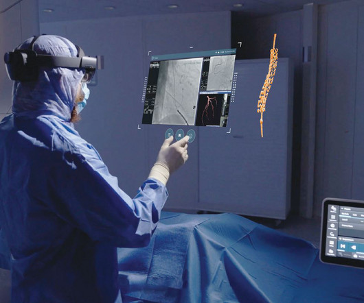
Image-guided therapy is a fast-evolving space that leverages advanced imaging and visualization solutions to make it possible to treat many conditions, from cancer care to cardiovascular disease and more, with little to no surgical incisions.
Radiology is a medical imaging procedure that uses ionizing electromagnetic radiation to create images of bones, organs, and soft tissues to diagnose a patient’s symptoms, disease, or conditions. It includes techniques like X-rays, CT scans, MRIs, ultrasounds, and fluoroscopy.
AP Chest X-ray demonstrating placement of 3 MitraClips over the tricuspid valve. A: AP chest X-ray demonstrating placement of 3 MitraClips over the tricuspid valve (yellow arrow) B: Coronal chest CT showing 3 MitraClips over the tricuspid valve in the right atrioventricular septum (red arrow). Name the cardiac device.
Ultrasound model predicts liver disease progression. Commercially Available Chest Radiograph AI Tools for Detecting Airspace Disease, Pneumothorax, and Pleural Effusion. Increased Metabolic Activity of the Thymus and Lymph Nodes in Pediatric Oncology Patients After Coronavirus Disease 2019 Vaccination.
We organize all of the trending information in your field so you don't have to. Join 5,000 users and stay up to date on the latest articles your peers are reading.
You know about us, now we want to get to know you!
Let's personalize your content
Let's get even more personalized
We recognize your account from another site in our network, please click 'Send Email' below to continue with verifying your account and setting a password.


Let's personalize your content