This site uses cookies to improve your experience. To help us insure we adhere to various privacy regulations, please select your country/region of residence. If you do not select a country, we will assume you are from the United States. Select your Cookie Settings or view our Privacy Policy and Terms of Use.
Cookie Settings
Cookies and similar technologies are used on this website for proper function of the website, for tracking performance analytics and for marketing purposes. We and some of our third-party providers may use cookie data for various purposes. Please review the cookie settings below and choose your preference.
Used for the proper function of the website
Used for monitoring website traffic and interactions
Cookie Settings
Cookies and similar technologies are used on this website for proper function of the website, for tracking performance analytics and for marketing purposes. We and some of our third-party providers may use cookie data for various purposes. Please review the cookie settings below and choose your preference.
Strictly Necessary: Used for the proper function of the website
Performance/Analytics: Used for monitoring website traffic and interactions
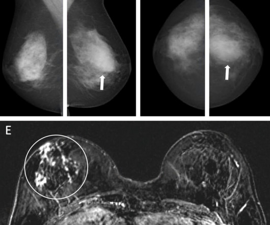
Using AI with mammography can help select women at high risk of breast cancer for supplemental MRI, according to research published February 4 in Radiology. Women at high risk of breast cancer, such as those with a personal history or family history of the disease or those with dense breasts, may be recommended for supplemental imaging.
The study results suggest that the model, called TotalSegmentator MRI, could improve radiology department workload, said senior author Jakob Wasserthal, PhD, of University Hospital Basel in Switzerland. To this end, D'Antonoli and colleagues built TotalSegmentator MRI, which they based on nnU-Net. for 13 anatomic structures).
X-ray and ultrasound machines were badly damaged in a rocket attack on Ukraine's largest children's hospital on July 8, according to radiologist Stanislav Rebenkov, MD. Rebenkov has been head of radiology at Ohmatdyt Children's Hospital since 2020. Broken glass and damaged equipment are found throughout the hospital.
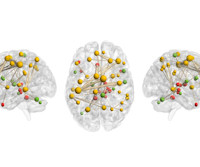
Functional MRI (fMRI) has revealed a brain circuit for creativity, researchers from Mass General Brigham in Boston have reported. The neuroanatomical origins of creativity have long been of interest to researchers, and may also help clinicians better understand symptoms among patients with various brain diseases.
Researchers using quantitative susceptibility mapping (QSM) MRI have discovered evidence of the long-term effects of severe COVID-19 on the brain. reported brain imaging findings on MRI in COVID-19 survivors who had been hospitalized. But brainstem involvement identified on MRI after COVID-19 recovery has remained inconclusive.
So as to report more CT and MRI, radiologists stopped doing hands-on ultrasound and fluoroscopy. Many diagnostic radiologists became pure CT and MRI specialists. But the pressure kept building, and the number of CT and MRI scans grew by 20% annually in my hospital. So radiologists started dropping duties.
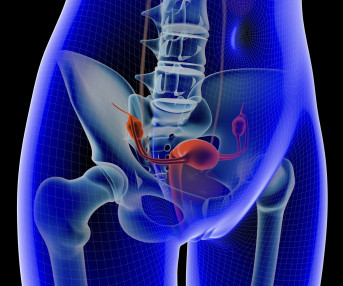
The findings could help clinicians tailor biopsy recommendations for men with low- to intermediate risk of the disease, according to the authors. Standard clinical practice has shifted from conducting systematic prostate biopsies without MRI guidance to using MRI-guided biopsies," the team wrote.
Middlebrooks' research interest consists of using ultrahigh-field, 7-tesla MRI to plot brain microstructure and develop surgical treatment of brain tumors, epilepsy, and neurodegenerative and movement disorders such as Parkinson's disease, essential tremor, and dystonia. Middlebrooks, MD, of the Mayo Clinic in Jacksonville, FL.
A large study, needed for FDA clearance, demonstrated that the use of icobrain aria significantly increases the accuracy of ARIA assessments by radiologists and hence allows for safer use of new amyloid-beta targeting therapies for Alzheimer’s disease patients. Example of how ARIA can present itself on a brain MRI scan.
PET/MRI imaging shows promise in diagnosing fevers or inflammation of unknown origin and may have advantages over PET/CT, according to a study published January 3 in the European Journal of Radiology. They evaluated the sensitivity, specificity, and predictive values of the PET/MRI findings in relation to the final diagnosis.
Gastrointestinal imagers should use high-quality MR enterography for complex inflammatory bowel disease (IBD) cases that can't be dealt with using intestinal ultrasound, even though both tests contribute to overall assessment, said an expert leading an advanced course on imaging Crohn's disease at ECR 2025. "MR
F-18 FDG-PET/MRI scans without the use of a contrast agent should be the top choice for imaging children suffering from lymphoma, according to research presented recently at RSNA in Chicago. Lymphomas represent approximately 15% of all tumor diseases in children and imaging plays a key role in tumor staging to plan treatments.
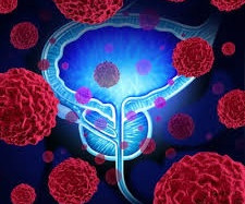
PET/CT imaging with a new radiotracer (F-18 PSMA-1007) is superior to MRI prior to surgery in men with intermediate-risk and high-risk prostate cancer, according to a study published on July 1 in JAMA Oncology.
Combining ultrasound with MRI could help better differentiate between benign and malignant breast nonmass-like lesions initially detected on ultrasound, a study published October 12 in Ultrasound in Medicine & Biology found. Ultrasound and MRI have their tradeoffs in supplemental breast imaging. 0.849 0.901 Specificity 52.3%
Massimo Mischi, PhD, of the Eindhoven University of Technology in the Netherlands, described the use of ultrasound contrast agents with AI and an imaging technique known as contrast-enhanced ultrasound dispersion imaging (CUDI) to produce 3D and 4D datasets that are as good as MRI imaging.
tesla MRI AI body composition analysis Cardiac PET Cryo/thermoablation CT colonography Genicular artery embolization Hyperpolarized xenon-129 MRI PET/MRI Photon-counting CT Radiomics Theranostics Whole-body MRI screening Image of the Year 3D PET/MR image.
A machine learning (ML) model including both coronary CT angiography (CCTA) and stress cardiac MRI data can accurately predict major adverse cardiovascular events (MACE), suggest findings published January 14 in Radiology. The retrospective study included data collected between 2008 and 2020 from 2,210 patients who completed cardiac MRI.
MRI reveals the negative effects of preeclampsia on pregnant women's hearts and on the brains of their fetuses, according to research published in the April issue of Hypertension. The results highlight MRI's benefits as a prenatal assessment tool, wrote a group led by Megan Hall, MD, of St Thomas' Hospital in London, U.K. "As
Researchers have found that MRI reveals functional brain alterations that are associated with major depression, according to a study published February 19 in JAMA Network Open. Those who had been identified with Hospital ICD-10 diagnosis codes F32 and F33 and antidepressant use had the most alterations in these measures. "We
Abbreviated breast MRI is not only effective for screening among high-risk women but also useful for evaluating "pure" ductal carcinoma in situ (DCIS), according to two presentations delivered March 2 at ECR 2024. Bartholomew's Hospital in London. Sensitivity 100% 100% Specificity 94.7%
The models can also assess the risk for early recurrence and progression-free survival, noted a team led by Yixing Yu, MD, of the First Affiliated Hospital of Soochow University in Suzhou, China. VETC is a novel vascular pattern of HCC associated with poor prognosis and benefit of sorafenib treatment," the group wrote. "In
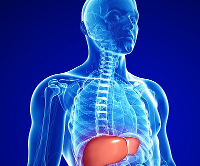
The MRI PDFF measure could improve patient care by giving clinicians a more accurate picture of liver disease, a team led by Tianyi Xia, MD, of Zhongda Hospital School of Medicine, Southeast University, in Nanjing, China. There are a variety of ways to assess liver fat content, but MRI PDFF has shown high diagnostic precision.
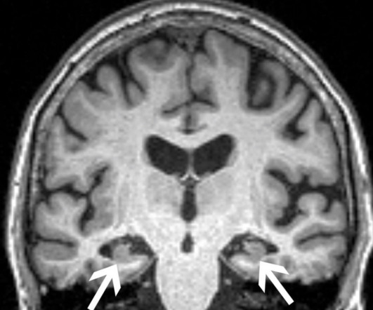
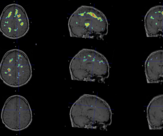
CHICAGO – Black patients receive fewer MRI scans for Alzheimer’s disease diagnosis and undergo imaging at an older age than other racial groups, according to research presented November 27 at RSNA 2023. A brain MRI in patient with mild cognitive impairment. Image courtesy of Joshua Wibecan, MD. in the Other group. “Our
A team led by study senior author William Palmer, MD, of Massachusetts General Hospital in Boston found that radiologists who had access to information on patients' symptoms when they interpreted MRI spine exams achieved "a near statistically perfect agreement with spine specialists."
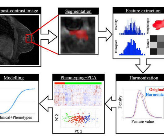
Time-dependent diffusion MRI-based microstructural mapping helps predict breast cancer molecular subtypes and treatment response in women with the disease, researchers have reported. The research included 408 women with breast cancer who underwent a time-dependent diffusion MRI exam between February 2021 and May 2023.
An MRI-based radiomics model shows potential for distinguishing low- from high-risk cases of ductal carcinoma in situ (DCIS), an early form of breast cancer, according to a study published April 1 in Radiology. Most women undergo surgery, yet only 26% of DCIS cases are subsequently identified as invasive disease, the authors noted.
Research presented at ECR 2024 suggests that diffusion-weighted MRI (DW-MRI) has value in ovarian cancer treatment planning. The Netherlands Cancer Institute, Leiden University Medical Center, and Catharina Ziekenhuis Teaching Hospital are collaborating on the multicenter MISSION trial. and in the interval group (AUC 0.83).
MR elastography (MRE) is an effective technique for noninvasive monitoring of liver stiffness -- a surrogate for fibrosis -- in children and young adults with autoimmune liver disease, researchers have reported. tesla MRI that included liver MRE as well as a liver biopsy within six months. years) who underwent an abdominal 1.5-tesla
OpenAI's GPT-4 AI model can utilize imaging reports to generate summaries of disease course in patients with complex glioblastoma, improving treatment planning and potentially even enhancing radiology workflows, according to research published July 23 in Radiology. No image data were transmitted to GPT-4.
Integrating radiology report features into a deep-learning (DL) algorithm improves the model's ability to identify brain lesions on MRI exams, researchers have reported. In addition, 2,655 brain MRI scans taken between January and December 2022 were used for external testing. The researchers extracted textual features (i.e.,
This study aims to provide a new evaluation method for diagnosing and treating TMT poisoning while shedding light on its underlying mechanisms, wrote first author Anqing Liu, of the Guangdong Province Hospital for Occupational Disease Prevention and Treatment. The study was published January 8 in Scientific Reports.
Centers for Disease Control and Prevention (CDC) acknowledges that “health disparities are preventable differences in the burden of disease, injury, violence, or opportunities.” Hospitals committed to addressing the root causes of healthcare disparities can start by addressing some of the following opportunities.
PET brain scans show persistent brain inflammation in patients with multiple sclerosis (MS), despite being treated with high-efficacy disease-modifying therapies, according to a recent study by researchers in Boston. This allows extended imaging acquisition times to measure TSPO as a biomarker, the team wrote.
Is there a signature biomarker for younger/early-onset Alzheimer’s Disease (EOAD)? Sporadic early-onset Alzheimer’s disease (EOAD) is rare and understudied. Still, these studies have included small samples and have not focused on developing an MRI biomarker. What Question Were You Investigating? What Were Your Findings?
Contrast-enhanced mammography (CEM) can locally stage lobular breast carcinomas and could serve as an alternative to breast MRI, according to research published February 12 in Clinical Radiology. A team led by Elisabetta Giannotti, MD, from Hospital NHS Foundation Trust in Cambridge, U.K., cm larger than those seen on MRI.
His research interests include using structural and functional MRI -- particularly ultrahigh-field, 7-tesla MRI -- to map brain microstructure and develop neurosurgical treatment of brain tumors, epilepsy, and neurodegenerative and movement disorders such as Parkinson's disease, essential tremor, and dystonia.
Arterial spin labeling MRI shows that alterations in women's cerebral blood flow begin during perimenopause and may be due to an increased burden of white-matter hyperintensities, researchers have reported. The group's findings were published October 18 in Stroke. "[Our]
Clinically significant extracardiac findings are common on CT and MRI imaging and are especially associated with exam indication and patient age, researchers have reported. Detecting and interpreting these findings is crucial for patient care, wrote a team led by Lukas Moser, MD, of University Hospital Zurich in Switzerland.
MRI is the imaging gold standard for diagnosis, yet identifying the disease using this method remains challenging, the researchers wrote. In all three cases, gadolinium-enhanced MRI scans did not show abnormalities. F-18 FDG-PET and MRI scans of three patients. (A.1-B.1) In our outpatient clinic follow-up (2 to 2.5
SINGAPORE -- The use of MRI for musculoskeletal (MSK) trauma imaging is becoming increasingly viable, according to a presentation delivered May 7 at the International Society of Magnetic Resonance in Imaging (ISMRM) meeting. MRI is underutilized in trauma imaging and often falls short of its potential," said presenter Prof.
In recent years, researchers have gained a deeper understanding of the complexity and variability of cancer and how each individuals body, molecular structure, and risk factors interplay with the disease. As an example, consider lung cancer, which is usually detected in the diseases advanced stages when treatment options are less effective.
Researchers led by Rui Tang from Peking University Third Hospital in Beijing, China, found that their AI system can accurately identify standard planes and perform automatic tissue segmentation. While ultrasound is a go-to method for assessing shoulder joint diseases, it is a user-dependent modality.
Food and Drug Administration (FDA) clearance to market its NM-101 AI platform for the analysis of neuromelanin-sensitive MRI scans. NM-101 is a cloud-based analysis platform designed to integrate into existing workflows at hospitals and imaging centers.
Researchers from Zurich have developed a cumulative clinical risk (CCR) score that may provide an early predictive marker for long-term brain alterations and cognitive impairments in patients with congenital heart disease (CHD). Figure courtesy of Melanie Ehrler, PhD, et al. moderate to strong effect sizes).
We organize all of the trending information in your field so you don't have to. Join 5,000 users and stay up to date on the latest articles your peers are reading.
You know about us, now we want to get to know you!
Let's personalize your content
Let's get even more personalized
We recognize your account from another site in our network, please click 'Send Email' below to continue with verifying your account and setting a password.


Let's personalize your content