This site uses cookies to improve your experience. To help us insure we adhere to various privacy regulations, please select your country/region of residence. If you do not select a country, we will assume you are from the United States. Select your Cookie Settings or view our Privacy Policy and Terms of Use.
Cookie Settings
Cookies and similar technologies are used on this website for proper function of the website, for tracking performance analytics and for marketing purposes. We and some of our third-party providers may use cookie data for various purposes. Please review the cookie settings below and choose your preference.
Used for the proper function of the website
Used for monitoring website traffic and interactions
Cookie Settings
Cookies and similar technologies are used on this website for proper function of the website, for tracking performance analytics and for marketing purposes. We and some of our third-party providers may use cookie data for various purposes. Please review the cookie settings below and choose your preference.
Strictly Necessary: Used for the proper function of the website
Performance/Analytics: Used for monitoring website traffic and interactions
The way physicians identify illness is changing due to advances in medical imaging, which make early diagnosis quicker, more precise, and less invasive. As one of El Pasos top radiology centers, Professional Radiology is dedicated to offering cutting-edge imaging services that aid in the early detection of illnesses.
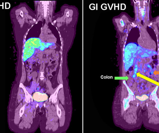
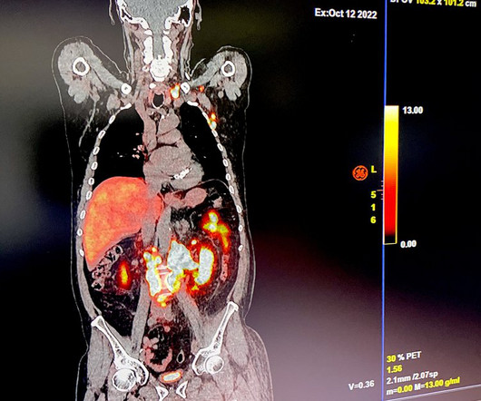
F-18 fluorothymidine (FLT) PET can identify early acute gastrointestinal graft versus host disease (GVHD) after patients undergo bone marrow transplants, according to a study published December 13 in Radiology: Imaging Cancer. F-18 FLT-PET imaging was performed on day 28. F-18 FLT-PET imaging of gastrointestinal GVHD.
A Swiss research team has developed and tested an AI model that automatically segments anatomic structures on MR images independent of sequence, according to a study published February 18 in Radiology. Images and caption courtesy of the RSNA. The team then assessed the model's performance using Dice scoring.
When it comes to medical imaging, radiology is what most often comes to mind, and for good reason. A large percentage of medical imaging created by most hospitals tends to come from the radiology department. In most cases, medical images and scans are placed into an electronic medical record (EMR) as a link. In the U.S.
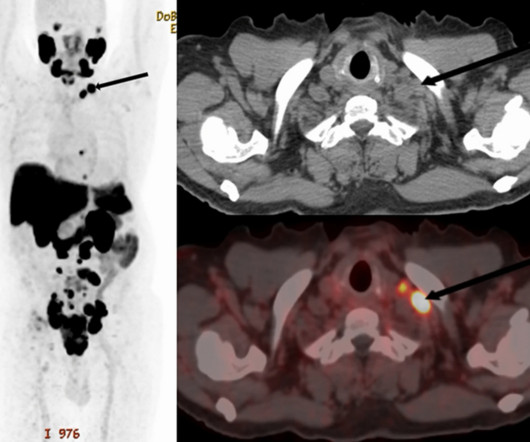
PET/CT scans with an experimental prostate-specific membrane antigen (PSMA) imaging agent can identify supraclavicular nodal metastasis in newly diagnosed prostate cancer patients, researchers have reported. For the analysis, the researchers included 240 patients who underwent scans for primary staging of newly diagnosed disease.
TORONTO -- AuntMinnie.com spoke with prostate cancer imaging expert Phillip Kuo, MD, PhD, of the City of Hope in Duarte, CA, at the Society of Nuclear Medicine and Molecular Imaging annual meeting about new developments in PET/CT imaging in patients on Lu-177 PSMA-617 therapy. last year for Alzheimer's disease.
However, mpMRI misses about 10% of cases, typically in patients with lower-grade disease and in patients with cribriform pattern disease, a subtype much more likely to recur after surgery or radiation therapy, he noted. Hybrid PET/MRI scanners were introduced about 15 years ago to leverage the advantages of both methods.
MRI illuminates the structural and functional organization of the brain -- thus helping clinicians predict the progression of Parkinson's disease in patients in its early stages, researchers have found. Parkinson's disease can manifest as tremors, slowness of movement, or rigidity, and its symptoms worsen over time, according to the RSNA.
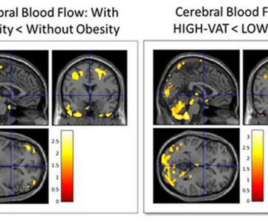
CHICAGO -- Characterizing an individual's type of body fat using body MRI can help predict Alzheimer's disease risk up to 20 years before symptoms manifest, according to research results presented December 2 at the RSNA meeting. Images courtesy of the RSNA.
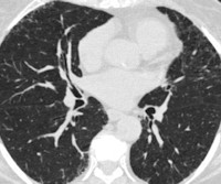
CT imaging shows that severe acute respiratory disease events can be caused by quantitative interstitial abnormalities (QIA) -- that is, small irregularities that don't necessarily meet diagnostic criteria for advanced pulmonary diseases but show up on CT exams over time, a study published April 30 in Radiology has reported.
PET imaging using a newly developed radiotracer has identified different patterns of brain tau pathology over time in early-onset versus late-onset Alzheimer’s disease patients, according to a study published February 1 in the Journal of Nuclear Medicine. Image courtesy of the Journal of Nuclear Medicine.
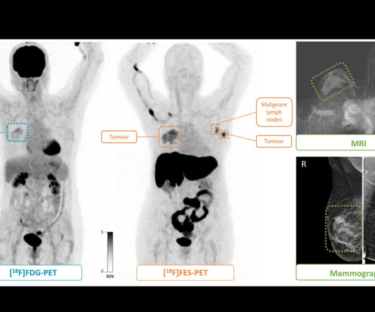
Conversely, F-18 FES-PET is a noninvasive, whole-body molecular imaging technique approved in 2020 that captures both spatial (differences based on location), and, when ER-positive patients are imaged at multiple time points, temporal heterogeneity (how tumors change over time), the authors noted. A visual abstract of the analysis.
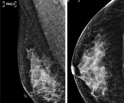
Benign breast disease diagnosed through percutaneous biopsy increases the overall risk of developing breast cancer, according to research published December 13 in JAMA Surgery. Benign breast disease makes up about 75% of breast biopsy diagnoses. The study included data from 4,819 women with a median age of 51.
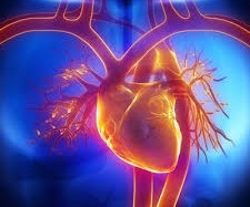
PET imaging with nitrogen-13 (N-13) ammonia radiotracer can help predict major adverse cardiac events in patients with ischemic heart disease (IHD), according to a study published May 30 in Radiology: Cardiothoracic Imaging. Patients underwent the scans between January 2017 and January 2021. The full study is available here.
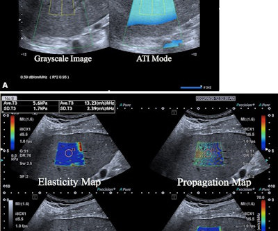
A predictive model combining multiple ultrasonic parameters could discriminate between different liver diseases in a study published March 19 in Radiology. These findings are important for noninvasive assessment of the MAFLD disease process,” the Liu team wrote. A 1-cm region of interest is placed within the sampling box of each image.
Deuterium metabolic imaging (DMI) can reveal impaired brain glucose metabolism in patients with Alzheimer’s disease, according to a study published July 16 in Radiology. DMI is an emerging method for imaging tissue metabolism based on MR spectroscopic detection of deuterium-labeled compounds, they explained.
Machine learning models could help create a more standardized, reproducible and efficient way of grading Crohn’s disease severity in the small bowel based on CT imaging.
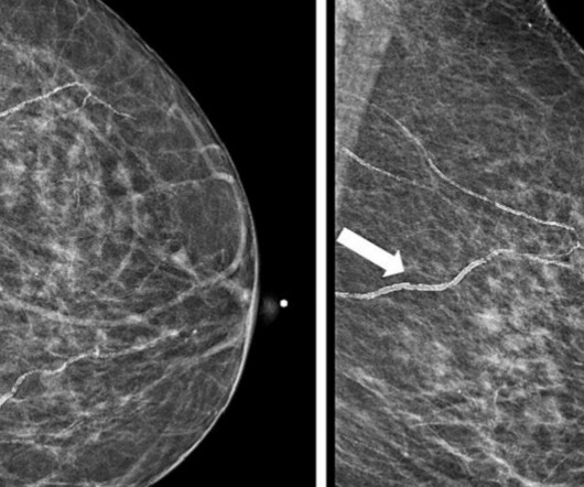
Postoperative MRI surveillance appears to lower the odds of advanced second breast cancer in women with a personal history of the disease, researchers have reported. "In Images in a 40-year-old woman who underwent breast-conserving surgery for left breast cancer and a surveillance breast MRI examination 25 months after surgery. (A)
ChatGPT-4 outperformed human clinicians in determining pretest and post-test disease probability after a negative test result involving chest radiographs and mammograms, according to a research letter published December 11 in JAMA Network Open. They compared its performance with a survey of 553 human clinicians from various specialties.
Imaging plays an essential role in addressing hepatic steatosis. Yet, radiologists have had almost zero involvement in shaping medical knowledge about the disease.
The percentage of lung fibrosis quantified on CT pulmonary angiograms (CTPA) by an AI model is associated with increased risk of mortality -- and boosts clinicians' ability to predict the survival of lung disease patients, researchers have found. Images courtesy of the RSNA. "[We The results were published February 6 in Radiology.
The use of AI in thoracic imaging has begun to demonstrate "cumulative evidence of effectiveness," but more testing and research are needed to determine its feasibility for this application, according to a commentary published February 25 in Radiology. van Beek, MD, of the University of Edinburgh in the U.K. in an accompanying editorial.
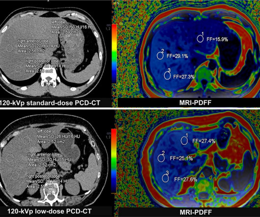
Photon-counting CT may serve as an alternative to MRI for assessing liver fat in patients with fatty liver disease, according to an article published September 24 in Radiology. The disease affects up to 24% of the population in the U.S. The disease affects up to 24% of the population in the U.S. Image courtesy of the RSNA.
In a recent PET study with unexpected results, patients infected with herpes had fewer signs of brain deposits associated with Alzheimers disease than uninfected patients, a group in France has reported. The study was published January 18 in Scientific Reports. The median age of the group was 74 years old. The full study is available here.
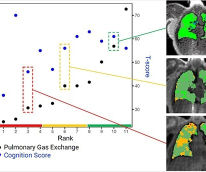
Image courtesy of RSNA. It's also a possibility that the disease mechanism that impairs pulmonary gas exchange also leads to higher brain perfusion through downstream vascular injury in both lung and brain," Staab said. Gas exchange maps of three representative patients from a low, medium, and high pulmonary gas exchange.
Some people with neurologic diseases experience a new onset of creative behavior and show specific patterns of damage that align with our creativity circuit," Kutsche said in a statement released by Mass General Brigham. The findings were published February 13 in JAMA Network Open.
Food and Drug Administration (FDA) has approved Eli Lilly's drug donanemab (Kisunla) for the treatment of early Alzheimer's disease, including mild cognitive impairment or mild dementia. Donanemab is a monoclonal antibody that targets and clears beta-amyloid protein in the brain.
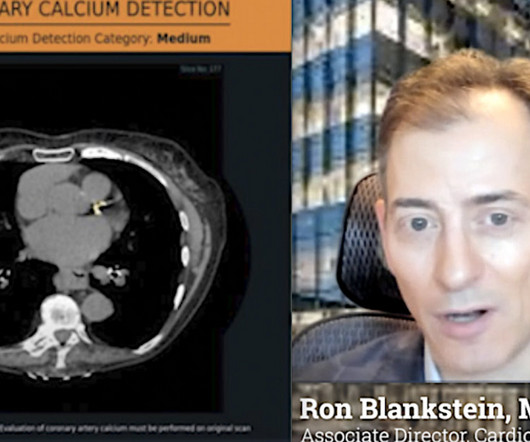
Ron Blankstein, MD, professor of radiology, Harvard Medical School, explains the use of artificial intelligence to detect heart disease in non-cardiac CT exams.
Understanding the mechanics of flow artifacts on CT or CT angiography (CTA) and how these artifacts are created is key to better disease diagnosis, according to a review published April 25 in RadioGraphics. These findings resolved (arrows in C) on a subsequent CT image (C) with a 70-second delay. Image and caption courtesy of the RSNA.
Our scans show where there is patchy ventilation in patients with lung disease, and show us which parts of the lung improve with treatment," Thelwall said in a statement released by the university. Image and caption courtesy of Newcastle University in Newcastle on Tyne, U.K.
Up to 30% of patients have metastatic disease by the time they are initially diagnosed with renal cell carcinoma, making accurate staging a critical element of treatment planning.
Preliminary studies suggest the tracer may also be effective in patients with earlier, more treatable stages of the disease, yet most of these studies have been retrospective and subject to a large risk of bias, the authors noted. Image and caption courtesy of the RSNA. All lesions had also been identified at mammography and MRI.
and radiology has learned much since then, according to experts who directly dealt with the diseases impact. While the pandemic affected medical operations across the country, the experts said that radiologists developed and honed their sense of resiliency as imaging was placed on the front lines.
Radiologist Jelle Barentsz, MD, PhD, of Radboud University in the Netherlands, emphasized that poor-quality MRI images are leading to prostate cancer overdiagnosis. We need to have a quality measurement of the imaging," he explained. "We We developed PI-QUAL which is, I think, the first MRI standard looking at image quality.
Functional MR (fMRI) imaging shows structural and functional alterations in the brains of people with opioid use disorder, according to a team of researchers from Yale School of Medicine in New Haven, CT. We observed widespread increases in global connectivity in individuals with [the disease]," Mehta said in a statement released by the RSNA.
PET, CT, and MRI use for assessing coronary artery disease increased in Medicare patients from 2018 to 2022, while SPECT and stress echocardiography declined over the same period, researchers have reported. Taken together, these findings call for efforts to address barriers impeding wider uptake of PET,” the group wrote.
This goes for women in all age groups and women who are Asian, Black, Hispanic, and Native American, according to the report written by Edward Hendrick, PhD, from the University of Colorado in Aurora and Debra Monticciolo, MD, from the Foundation for Imaging Research and Education in Temple, TX. We encourage women to get screened.
A new study suggests sildenafil, known by its brand name Viagra, can improve cerebrovascular function and minimize the risk of cognitive impairment caused by small vessel disease.
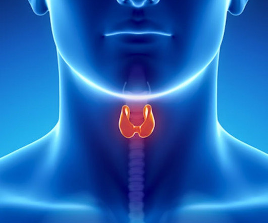
Outcomes over 10 years suggest that image-guided thermal ablation is a safe and effective option for patients with papillary thyroid carcinoma, according to a study published November 7 in JAMA Otolaryngology-Head & Neck Surgery. The 10-year disease-free survival was 93.9%. The mean age of patients was 45.8
ChatGPT would require further refinement before being used clinically to help educate patients about cardiac imaging, according to a study published May 23 in Clinical Imaging. Questions ranged from those on simple diagnostic subjects (“My doctor wants to run tests to diagnose coronary artery disease.
We organize all of the trending information in your field so you don't have to. Join 5,000 users and stay up to date on the latest articles your peers are reading.
You know about us, now we want to get to know you!
Let's personalize your content
Let's get even more personalized
We recognize your account from another site in our network, please click 'Send Email' below to continue with verifying your account and setting a password.


Let's personalize your content