This site uses cookies to improve your experience. To help us insure we adhere to various privacy regulations, please select your country/region of residence. If you do not select a country, we will assume you are from the United States. Select your Cookie Settings or view our Privacy Policy and Terms of Use.
Cookie Settings
Cookies and similar technologies are used on this website for proper function of the website, for tracking performance analytics and for marketing purposes. We and some of our third-party providers may use cookie data for various purposes. Please review the cookie settings below and choose your preference.
Used for the proper function of the website
Used for monitoring website traffic and interactions
Cookie Settings
Cookies and similar technologies are used on this website for proper function of the website, for tracking performance analytics and for marketing purposes. We and some of our third-party providers may use cookie data for various purposes. Please review the cookie settings below and choose your preference.
Strictly Necessary: Used for the proper function of the website
Performance/Analytics: Used for monitoring website traffic and interactions
Although the most common site of MBC is bone, there is currently no standardized imagingmodality that offers accurate assessment of bone treatment response, Masperi and Girlando explained. Data on radiation doses and the administration of contrast agents or radiopharmaceuticals were also collected.
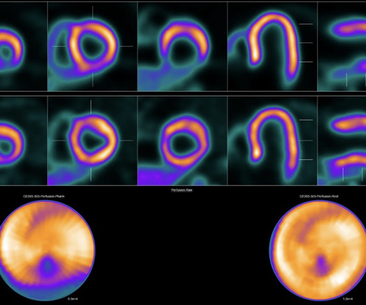
Strategies to increase the use of cardiac PET myocardial perfusion imaging (MPI) in the U.S. Cardiac PET MPI has emerged as a key tool for diagnosing and managing patients with cardiovascular diseases, especially coronary artery disease, yet overall it remains underutilized in the U.S., the authors noted.
PEM is one molecular breast imaging method that has shown promise in lowering the number of false-positive cases, owing to its superior specificity over MRI. However, PEM’s higher radiation dose has steered radiologists away from using the modality. On 100 sets of bilateral PEM images, 24 (96%) of 25 cancers were identified.
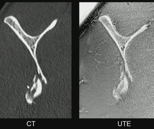
18F-flurpiridaz PET MPI obtained images at a lower radiation dose than 99Tc-SPECT MPI and performed similarly in both obese and non-obese patients. “Due This can result in inferior image quality and diagnostic performance despite requiring a higher dose of radiation.”
Ultrasound proponents will have the opportunity to present and see the modality’s versatility in helping detect and diagnose pathologies such as liver cancer, breast cancer, and thyroid cancer. They can also highlight the resulting benefits that are possible, of course, without the ionizing radiation of some other imagingmodalities.
Even if some private urology, radiation oncology practices, or radiologist groups are building the ability to perform theranostics, experts are cautious about patient management, radiation safety, and the risk of unnecessary imaging. However, few freestanding theranostics centers exist today. Morris continued in the JNM.
Fritz moderated a session titled "Seeing the Unseen: MRI in Traumatic Musculoskeletal Disease." X-ray and CT imaging are the go-to exams for trauma, Fritz conceded, but MRI is gaining ground as an effective modality for MSK trauma applications, in part because it does not expose patients to radiation and because it can image soft tissue well.
mtaschetta-millane Tue, 07/02/2024 - 09:53 July 2, 2024 — A new editorial paper was published in Oncoscience ( Volume 11 ) on May 20, 2024, entitled, “ Deep learning-assisted lesion segmentation in PET/CT imaging: A feasibility study for salvage radiation therapy in prostate cancer.” In this new editorial, researchers Richard L.J.
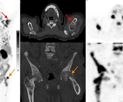
milla1cf Tue, 08/15/2023 - 16:28 August 15, 2023 — In elderly patients with suspected prostate cancer, PSMA PET/CT can diagnose advanced disease and aid in therapy selection without the need for a biopsy. Compared with patients who did have a pre-imaging biopsy, these patients were older with worse clinical status and had higher PSA levels.
While neuromelanin was first described in the 19 th century, researchers have elucidated its possible role as a biomarker for neurological disorders such as Parkinson’s disease in the last two decades. Cassidy, “it allows us to obtain potentially useful adjunctive information about many other diseases such as Alzheimer’s disease, and PTSD.”
Since almost half of the screening population has dense breasts, many of these patients require additional breast imaging, often with MRI , after mammography. Low-dose positron emission mammography (PEM) is a novel molecular imaging technique that provides improved diagnostic performance at a radiation dose comparable to that of mammography.
i Thankfully, the field of breast screening is not static, and advancements are revolutionizing how we detect and diagnose the disease. For dense-breasted patients requiring supplemental imaging, MRI remains a valuable option that is not limited by breast density and is shown to be more sensitive than mammography at finding breast cancer.
For many, machine learning's potential is more than just talk; the power of well-trained, image-augmented AI has already been demonstrated as a useful tool for quality control, disease tracking, tumor segmentation, and making prognosis predictions.
Early Disease Detection : Radiological imaging detects diseases in their earliest stages, significantly improving the chances of successful treatment and patient outcomes.
Discuss how this approach delivers radiation with exceptional precision to cancer cells. Advances in Cardiac Imaging: Precision in Heart Disease Diagnosis: Highlight recent breakthroughs in cardiac imaging within nuclear medicine.
Advanced algorithms can analyze images more quickly and accurately, aiding in early diagnosis and treatment planning. 3D and 4D Imaging : The development of 3D and 4D imaging techniques provides a more comprehensive and dynamic view of internal structures, allowing for improved anatomical understanding and better treatment planning.
The balance of dose and image quality is even more important in pediatric medical imaging. Not only are children more radiosensitive than adults (the cancer risk per unit dose of ionizing radiation is higher), but children also have a longer expected lifetime, which puts them at greater risk of cancer following radiation exposure.(1)
Teleradiology & Radiology data for artificial intelligence (AI) Introduction: Embark on a journey into the world of medical imaging as we unravel the distinctions between two powerful diagnostic tools—Computed Tomography (CT) scans and Positron Emission Tomography (PET) scans.
Introduction to Radiology : Radiology is a branch of medicine that uses medical imaging techniques to diagnose and treat diseases and injuries. It includes various imagingmodalities such as X-rays, CT scans, MRIs, ultrasounds, and nuclear medicine.
Digital Transformation : Traditional film-based radiography is being replaced by digital imaging systems, which offer higher resolution, more efficient data management, and the ability to share images electronically.
Advanced ImagingModalities: Unveiling the Microscopic World: Explore the advancements in imagingmodalities such as high-resolution MRI and diffusion tensor imaging. Neuroimaging in Neurodegenerative Diseases: Early Detection and Monitoring: Explore the role of neuroimaging in neurodegenerative diseases.
Medical imaging is a crucial tool in modern healthcare, providing detailed visuals of the human body’s internal structures and helping in the accurate diagnosis and treatment of various conditions. Magnetic Resonance Imaging (MRI) MRI stands for Magnetic Resonance Imaging. X-rays are fast, painless, and commonly used.
They are used in the diagnosis of bone fractures, lung diseases, dental issues, and various other medical conditions. Advanced ImagingModalities: X-ray technology has expanded beyond conventional radiography. These advanced imagingmodalities offer unparalleled insights into anatomy and pathology.
It can help reduce the symptoms of tremor, slowness, stiffness, and walking problems caused by Parkinson’s disease, dystonia, or essential tremor. Preoperative brain imaging (usually MR imaging) is used principally for the selection of those patients with PD who are candidates for DBS intervention (bilateral GPi or STN DBS).
There is no history or physical exam feature that rules out the disease Lactate elevation is a late finding in SBO. Examples: adhesions, neoplasms, inflammatory disease (i.e. There is no history or physical exam feature that rules out the disease Lactate elevation is a late finding in SBO. Read more
This technology can also offer improved image clarity, allowing radiologists to discern individual organs, tissue, and structures and reduce the risk of misdiagnosis. These technologies can help radiologists and medical professionals obtain detailed images of the entire body or specific organs to detect diseases or injuries more effectively.
A group led by researchers at the University of Hong Kong in China found the approach detected 12% more disease in patients undergoing staging than CT or MRI and resulted in a change in treatment in a significant number of cases. The results validate the emerging technique, the group noted.
What if X-ray imaging, the most prevalent and accessible imagingmodality in the world, could provide the information needed for diagnosis? The study could lead to radiographs providing an early window into disease manifestations that are currently undetected, thus leading to an earlier diagnosis of IPF.
The management of prostate cancer can be said to be a complex issue as there are a lot of challenges involved in accurate staging and predicting the speed of disease progression. MRI Imaging of Prostate was started sometime during the mid-1980’s. Proliferation of cells through the membrane causes prostate cancer.
3) The British Röntgen Society (the first radiology society) was founded in 1897, and many further studies on X-ray usage and the effects of radiation were performed over the following years. (3) 15) Radiologists needed a common means for sharing images. 3) This is what is known as tomography. Radiology 1963;80:273–275 Lindsay, R.
MRI before and after a gadolinium-based contrast agent is the preferred imagingmodality for evaluating brain tumors. Treatment varies by tumor type and often includes a combination of surgery, chemotherapy, and radiation. Diagnosis requires tumor biopsy with consideration of histopathological and molecular characteristics.
Newborns' livers can be affected by a variety of congenital and acquired diseases, and imaging plays an important role in the workup and management of these, according to a study published November 7 in RadioGraphics. Although they are rare, various congenital and acquired diseases can affect the neonatal liver," the group wrote.
We organize all of the trending information in your field so you don't have to. Join 5,000 users and stay up to date on the latest articles your peers are reading.
You know about us, now we want to get to know you!
Let's personalize your content
Let's get even more personalized
We recognize your account from another site in our network, please click 'Send Email' below to continue with verifying your account and setting a password.


Let's personalize your content