This site uses cookies to improve your experience. To help us insure we adhere to various privacy regulations, please select your country/region of residence. If you do not select a country, we will assume you are from the United States. Select your Cookie Settings or view our Privacy Policy and Terms of Use.
Cookie Settings
Cookies and similar technologies are used on this website for proper function of the website, for tracking performance analytics and for marketing purposes. We and some of our third-party providers may use cookie data for various purposes. Please review the cookie settings below and choose your preference.
Used for the proper function of the website
Used for monitoring website traffic and interactions
Cookie Settings
Cookies and similar technologies are used on this website for proper function of the website, for tracking performance analytics and for marketing purposes. We and some of our third-party providers may use cookie data for various purposes. Please review the cookie settings below and choose your preference.
Strictly Necessary: Used for the proper function of the website
Performance/Analytics: Used for monitoring website traffic and interactions
Imaging was so important [for cardiac indications], that I decided to become a radiologist," he said. Together with our radiographers, I learned to scan cardiac patients and learned special anatomy from pediatric cardiologists and pediatric cardiac surgeons." years of follow-up compared with CT imaging (2.1% But he persevered.
A team led by Junqi Han, MD, from the Affiliated Hospital of Qingdao University in China found that its model combining data from mammography images, ultrasound images, and other characteristics performed well in predicting disease-free survival of breast cancer.
i Thankfully, the field of breast screening is not static, and advancements are revolutionizing how we detect and diagnose the disease. For dense-breasted patients requiring supplemental imaging, MRI remains a valuable option that is not limited by breast density and is shown to be more sensitive than mammography at finding breast cancer.
Advanced algorithms can analyze images more quickly and accurately, aiding in early diagnosis and treatment planning. 3D and 4D Imaging : The development of 3D and 4D imaging techniques provides a more comprehensive and dynamic view of internal structures, allowing for improved anatomical understanding and better treatment planning.
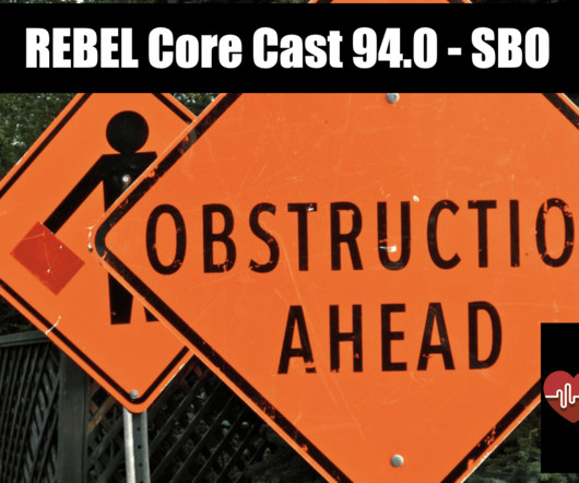
There is no history or physical exam feature that rules out the disease Lactate elevation is a late finding in SBO. Examples: adhesions, neoplasms, inflammatory disease (i.e. There is no history or physical exam feature that rules out the disease Lactate elevation is a late finding in SBO. Read more
The CPT code for Ultrasound Abdomen Complete includes the bowel and it is perfectly acceptable to incorporate views of the pylorus, ileocecal valve and appendix, the portions of the bowel where disease is likely to occur no matter what your age, as a part of your routine scanning protocol.
1 Imaging: Computed tomography (CT) is the recommended imagingmodality for evaluating orbital trauma. Following surgery, patients should begin antibiotic coverage of pathogens associated with endophthalmitis per recommendations provided by their institution’s infectious disease specialist. Imaging of orbital trauma.
What if X-ray imaging, the most prevalent and accessible imagingmodality in the world, could provide the information needed for diagnosis? The study could lead to radiographs providing an early window into disease manifestations that are currently undetected, thus leading to an earlier diagnosis of IPF.
after seeing the image. (2) Photoprint from radiograph by W.K. 3) In the early twentieth century, it was a common goal for investigators to try to find a way to separate the superimposed shadows that were recorded when a complex structure was shown on a radiograph. (3) 15) Radiologists needed a common means for sharing images.
Newborns' livers can be affected by a variety of congenital and acquired diseases, and imaging plays an important role in the workup and management of these, according to a study published November 7 in RadioGraphics. Imaging has [a key] role in the workup and management of many neonatal hepatic abnormalities. Ultrasound.
3] To identify a causative vascular lesion, which may or may not be amenable or contraindicatory to thrombolysis Non-Contrast Head CT NCCT is usually the first imagingmodality obtained in the acute evaluation for stroke. Vascular Diseases of the Brain.” Radiographics. Within the thrombolysis window (<4.5
We organize all of the trending information in your field so you don't have to. Join 5,000 users and stay up to date on the latest articles your peers are reading.
You know about us, now we want to get to know you!
Let's personalize your content
Let's get even more personalized
We recognize your account from another site in our network, please click 'Send Email' below to continue with verifying your account and setting a password.


Let's personalize your content