This site uses cookies to improve your experience. To help us insure we adhere to various privacy regulations, please select your country/region of residence. If you do not select a country, we will assume you are from the United States. Select your Cookie Settings or view our Privacy Policy and Terms of Use.
Cookie Settings
Cookies and similar technologies are used on this website for proper function of the website, for tracking performance analytics and for marketing purposes. We and some of our third-party providers may use cookie data for various purposes. Please review the cookie settings below and choose your preference.
Used for the proper function of the website
Used for monitoring website traffic and interactions
Cookie Settings
Cookies and similar technologies are used on this website for proper function of the website, for tracking performance analytics and for marketing purposes. We and some of our third-party providers may use cookie data for various purposes. Please review the cookie settings below and choose your preference.
Strictly Necessary: Used for the proper function of the website
Performance/Analytics: Used for monitoring website traffic and interactions
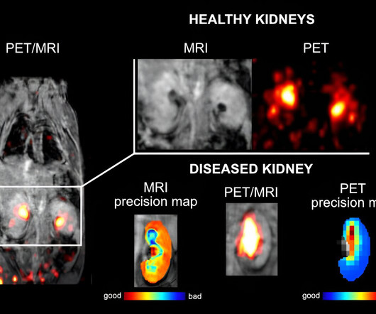
A research team from IOCB Prague, working in collaboration with the University of Tübingen, Germany, and the Faculty of Science, Charles University, has developed a new type of contrast agent that can be used in both magneticresonanceimaging (MRI) and positron emission tomography (PET).
For the detection of obstructive coronary artery disease (CAD), stress cardiovascular magneticresonanceimaging (MRI) demonstrated a sensitivity rate of 81 percent and a specificity rate of 86 percent, according to a meta-analysis of 64 studies and data from 74,470 patients with stable chest pain.
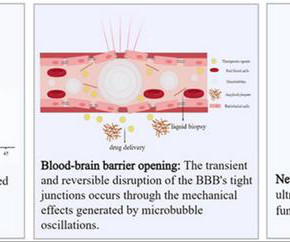
Food and Drug Administration has approved this technology for the treatment of essential tremor and Parkinson's disease, and its indications are expanding to include various intracranial diseases. High-intensity FUS is a safe and effective treatment for various intracranial diseases.
Since the advent of the magneticresonanceimaging (MRI) exam on human patients in the late 1970s, this innovation offered a multi-layered and noninvasive approach to the imaging of bodily organs, functions and the ability to diagnose disease.
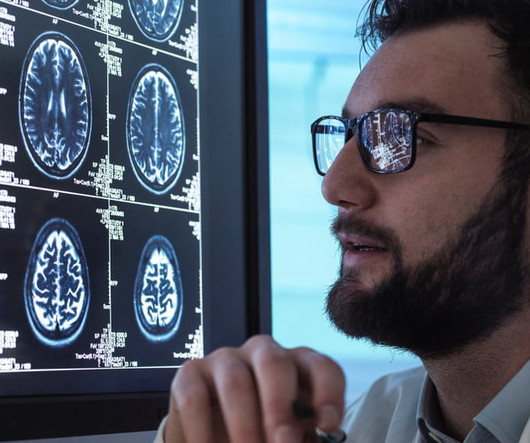
Is there a signature biomarker for younger/early-onset Alzheimer’s Disease (EOAD)? Sporadic early-onset Alzheimer’s disease (EOAD) is rare and understudied. Sporadic early-onset Alzheimer’s disease (EOAD) is rare and understudied. What Question Were You Investigating? What Were Your Findings? Alzheimer’s Dement.
PET-CT-Scan-Reporting-Service Introduction: MagneticResonanceImaging (MRI) is a complex and powerful diagnostic tool that requires mastery for effective use. Whether you are a healthcare professional or a student entering the field, this guide will help you navigate the intricate world of MagneticResonanceImaging.
While neuromelanin was first described in the 19 th century, researchers have elucidated its possible role as a biomarker for neurological disorders such as Parkinson’s disease in the last two decades. Cassidy, “it allows us to obtain potentially useful adjunctive information about many other diseases such as Alzheimer’s disease, and PTSD.”
Researchers at the University Medical Center Göttingen (UMG) and the Max Planck Institute for Multidisciplinary Sciences (MPI-NAT) have succeeded in visualizing the movement patterns of the internal speech muscles of a stuttering patient using real-time magneticresonanceimaging (MRI).
Reportedly receiving the first Current Procedural Terminology (CPT) III code from the American Medical Association (AMA) for artificial intelligence (AI)-enabled brain magneticresonanceimaging (MRI) software, Icometrix says its adjunctive quantification software can be utilized for diagnosis and assessment of conditions ranging from Alzheimer’s disease (..)
In certain cases, a new method can provide as much information from brain images taken with computed tomography as images captured with magneticresonanceimaging.
The artificial intelligence (AI)-powered Neuro Suite reportedly enables radiologists to access leading neurological AI algorithm solutions in the field, including the brain magneticresonanceimaging (MRI) segmentation capabilities of the Combinostics’ algorithm that can help differentiate degenerative pathologies such as Alzheimer’s disease and dementia. (..)
A large study, needed for FDA clearance, demonstrated that the use of icobrain aria significantly increases the accuracy of ARIA assessments by radiologists and hence allows for safer use of new amyloid-beta targeting therapies for Alzheimer’s disease patients.
milla1cf Fri, 10/20/2023 - 18:29 October 20, 2023 — In certain cases, a new method can provide as much information from brain images taken with computed tomography (CT) as images captured with magneticresonanceimaging (MRI). About two percent of all people over the age of 65 are affected.
milla1cf Fri, 11/03/2023 - 12:22 November 3, 2023 — Guerbet , a global leader in medical imaging with more than 30 years of experience in MRI, announced today its schedule of activities at the 2023 Radiological Society of North America (RSNA) Scientific Assembly and Annual Meeting from November 26-30 in Chicago, IL.
milla1cf Tue, 08/08/2023 - 20:34 August 8, 2023 — Fresenius Kabi announced it has launched Gadobutrol Injection, a generic substitute for the contrast agent Gadavist, which is used in magneticresonanceimaging (MRI) procedures. To assess the presence and extent of malignant breast disease in adult patients.
Magneticresonanceimaging (MRI) machines can clearly view non-bony parts of the body—soft tissue such as the brain, muscles and ligaments—as well as detect tumors, making it possible to diagnose many diseases and other conditions.
Siemens Healthineers and UH will look to also advance the treatment of patients with Alzheimer’s disease and use theranostics—combining the approaches of diagnostics and therapeutics—to treat patients with advanced forms of certain cancers, as well as develop new magneticresonance ( MR ) technologies. tesla and 3T scanners.
milla1cf Thu, 07/06/2023 - 22:19 July 7, 2023 — Bayer , a global leader in radiology, has initiated the Phase III clinical development program called QUANTI , aiming to evaluate the safety and efficacy of gadoquatrane, an investigational extracellular macrocyclic gadolinium-based contrast agent (GBCA) for use in magneticresonanceimaging ( MRI ).
1,2 "The final approval of gadopiclenol by the European Commission marks a significant milestone in the field of diagnostic imaging. 5 Gadolinium is a rare earth metal that has unique magnetic properties that make it useful for MRI imaging.
In today’s post, we hope to help you: Better understand the disease Know the approaches to screening See the available treatment options Patients may become confused after receiving a diagnosis due to the different types of prostate cancer.
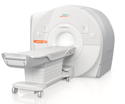
3 Tesla (3T) magneticresonanceimaging whole-body scanner. It possesses the strongest-ever gradient system for a clinically released whole-body MR scanner, making smaller structures in the body visible, and capturing images faster than previous MR scanners. Additionally, the Magnetom Cima.X
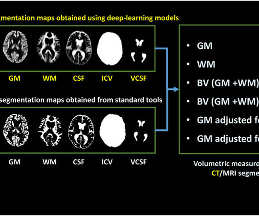
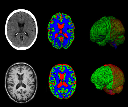
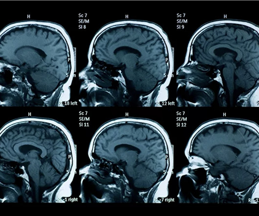
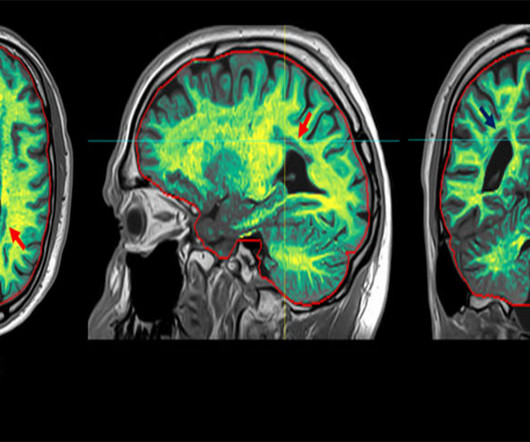
Accurate measurement of the volume and distribution of different tissue types in the brain is important for diagnosing brain disease. However, visual assessments of MR images are often subjective, and lack the necessary accuracy while measuring tissue volume from 2D image slices is virtually impossible.
milla1cf Thu, 01/04/2024 - 10:47 January 4, 2024 — Diagnosing cancer today involves using chemical “contrast agents” to improve the accuracy of medical imaging processes such as X-rays as well as computed tomography (CT) and magneticresonanceimaging (MRI) scans.
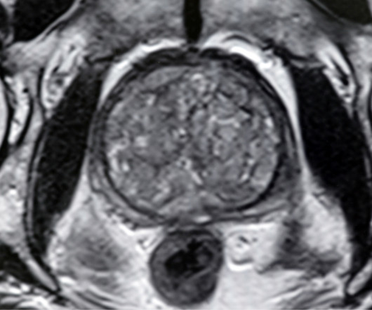
Understanding Prostate MRI Prostate MRI (MagneticResonanceImaging) is an advanced imaging technique used to visualize the prostate gland in high detail. Monitoring : For men on active surveillance for low-risk prostate cancer, MRI can be used to monitor disease progression, reducing the need for repeated biopsies.
The world of medical imaging is marking a significant milestone in 2023: the 50th anniversary of magneticresonanceimaging (MRI). It assists in identifying diseases related to spine lesions, tumors, and stroke impacting the area of blood vessels and brain.
The combination of Philips’ MR 7700 multi-nuclei scanner with the FDA-approved Xenoview hyperpolarized Xenon magneticresonanceimaging (MRI) contrast agent may facilitate earlier diagnosis and intervention for patients with obstructive lung diseases.
a second-generation clinical 7-tesla magneticresonanceimaging scanner that dramatically advances the capabilities of 7T MR scanning with new hardware and XA60A software. is the successor to the MAGNETOM Terra , which debuted in 2017 as the world’s first 7T clinical MR scanner, enabling extremely high-resolution imaging. “The
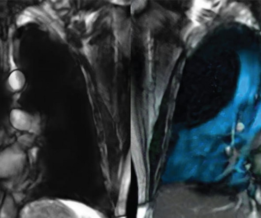
The overall purpose of this work is to quantify radiomics within Venous Malformations (VMs) and build disease progression and treatment prediction models. The quantification of radiomics necessitates the VM lesion segmentation. Therefore, the goal is to develop a fully automated algorithm to segment VMs in multiple body regions.
milla1cf Fri, 02/09/2024 - 09:41 February 9, 2024 — Multiple sclerosis (MS) is a neurological disease that usually leads to permanent disabilities. One key feature of the disease is that it causes the patient’s own immune system to attack and destroy the myelin sheaths in the central nervous system. It affects around 2.9
Furthermore, this partnership perfectly aligns with our objective of extending the reach of XENOVIEW MRI, to provide tools to physicians that treat patients suffering from chronic lung disease.” Susan Wood , Ph.D., CEO of VIDA, said: “VIDA and Polarean are natural partners.
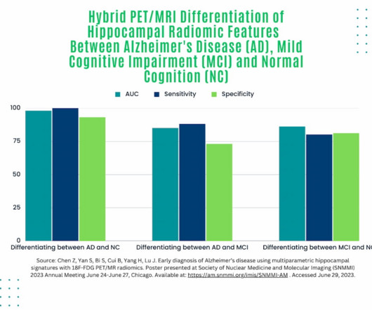
Employing a hybrid positron emission tomography (PET)/magneticresonanceimaging (MRI) model to assess predictive features of Alzheimer’s disease (AD), researchers noted a 100 percent sensitivity rate and a 93 percent sensitivity rate for distinguishing between AD and normal cognition, according to a study presented at the recent Society of Nuclear (..)
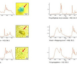
None of the individuals had died from neurological disease. Noninvasive imaging technologies, such as magneticresonanceimaging, are needed to overcome the current limitations in tissue analysis of different human organs and to improve the understanding of the health hazards of MPs,” the researchers concluded.
The availability of these offerings – including the MIM SurePlan and MIM Symphony families, MIM Maestro, MIM Encore, and more – is in alignment with GE HealthCare’s precision care strategy, which aims to deliver innovative digital solutions across care pathways for more precise, connected, and efficient care across disease states. "We
Evaluating the Tissue and Organs in the Chest Chest CT scans are are more detailed than x-rays, giving you more information about possible diseases or injury of your chest organs. CT scans also aid in surgical planning as well as monitoring the effectiveness of cancer treatments like chemo or radiation therapy.
There are several types of diagnostic imaging available today; each one used to visualize the internal structures of the body to assist doctors in diagnosis and treating various diseases and medical conditions. Professional Radiology strives to provide patients with accurate and compassionate imaging services.
On November 29, the company will present safety data for Elucirem in an abstract titled "Safety of gadopiclenol for magneticresonanceimaging (MRI): a pooled analysis of eight studies," it said. Guerbet plans to highlight its gadopiclenol contrast agent (Elucirem) for MRI at the upcoming RSNA meeting.
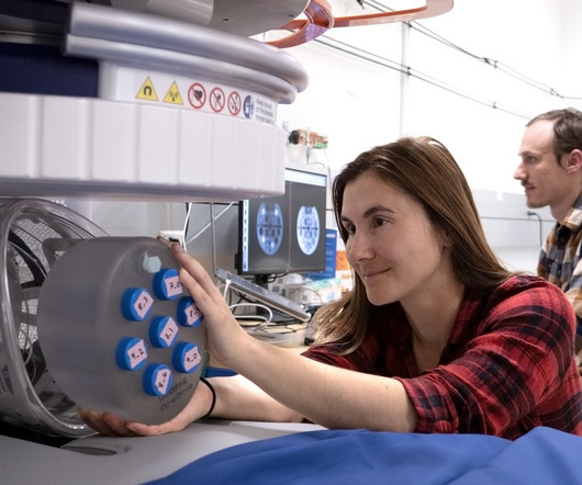
The Care Innovation Hub leverages the strengths of academia and industry to create, evaluate and translate novel technology into a clinical setting with the goals of advancing diagnosis and treatment of disease, improving hospital operations and driving more equitable access to care. tim.hodson Mon, 01/27/2025 - 10:11 Jan.
A 55-year-old woman with a history of end-stage kidney disease on peritoneal dialysis presents due to abdominal pain. Barium contrast X-ray CT scan Magneticresonanceimaging Ultrasound FOR THE RIGHT ANSWER CLICK ON THE ROSH REVIEW LOGO BELOW References Burkart JM, Bleyer A. End-stage renal disease. Post TW, ed.
55881 Ablation of prostate tissue, transurethral, using thermal ultrasound, including magneticresonanceimaging guidance for, and monitoring of, tissue ablation. PC-6.47 $525.63 $209.28 PC-14.56 $8,508.75 $470.97
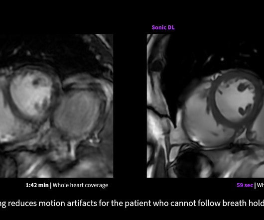
milla1cf Wed, 06/07/2023 - 19:51 June 6, 2023 — GE HealthCare announced the FDA clearance and launch of Sonic DL – a state-of-the-art deep learning-based technology designed to dramatically accelerate image acquisition in magneticresonanceimaging ( MRI ).
A team led by Stephane Loubrie, PhD, of the University of California San Diego in La Jolla found that information generated by a DWI-MRI technique called restriction spectrum imaging (RSI) showed significant difference between cancers and high-risk benign lesions compared with average-risk benign lesions.
Researchers have developed and substantiated an advanced magneticresonanceimaging (MRI) tool to reveal new structural insights into atherothrombosis, a long-term arterial vessel wall disease characterized by the build-up of lipid-rich and inflamed plaques.
We organize all of the trending information in your field so you don't have to. Join 5,000 users and stay up to date on the latest articles your peers are reading.
You know about us, now we want to get to know you!
Let's personalize your content
Let's get even more personalized
We recognize your account from another site in our network, please click 'Send Email' below to continue with verifying your account and setting a password.


Let's personalize your content