This site uses cookies to improve your experience. To help us insure we adhere to various privacy regulations, please select your country/region of residence. If you do not select a country, we will assume you are from the United States. Select your Cookie Settings or view our Privacy Policy and Terms of Use.
Cookie Settings
Cookies and similar technologies are used on this website for proper function of the website, for tracking performance analytics and for marketing purposes. We and some of our third-party providers may use cookie data for various purposes. Please review the cookie settings below and choose your preference.
Used for the proper function of the website
Used for monitoring website traffic and interactions
Cookie Settings
Cookies and similar technologies are used on this website for proper function of the website, for tracking performance analytics and for marketing purposes. We and some of our third-party providers may use cookie data for various purposes. Please review the cookie settings below and choose your preference.
Strictly Necessary: Used for the proper function of the website
Performance/Analytics: Used for monitoring website traffic and interactions
The voters also zeroed in on the ongoing shortage of radiologists as the Biggest Threat to Radiology. Importantly, we are gaining deeper insights into disease processes themselves, which enhances our ability to diagnose and potentially treat conditions that were previously beyond our reach." But these barriers aren't deal-breakers.
Opportunistic CT imaging data found on abdominal exams performed for other indications can be used to improve patientcare, according to a presentation delivered on September 12 at the International Society for Computed Tomography (ISCT) meeting.
Gastrointestinal imagers should use high-quality MR enterography for complex inflammatory bowel disease (IBD) cases that can't be dealt with using intestinal ultrasound, even though both tests contribute to overall assessment, said an expert leading an advanced course on imaging Crohn's disease at ECR 2025. "MR
A large study, needed for FDA clearance, demonstrated that the use of icobrain aria significantly increases the accuracy of ARIA assessments by radiologists and hence allows for safer use of new amyloid-beta targeting therapies for Alzheimer’s diseasepatients. icobrain aria was thoroughly evaluated in large reader studies.
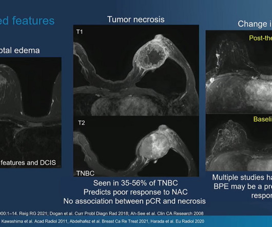
Five radiologists with between one and 30 years of experience generated the reports. 25% reported the extent of the disease. Additionally, no report in the impression included the pattern of response, 95% included the maximum extent of disease, and 40% included baseline size. 53% included preoperative MRI evaluation.
At the RSNA meeting, attendees will hear animated discussion on the opportunistic use of CT imaging, including, for example, how it can provide information on a patient's bone mineral density, catch pancreatic ductal adenocarcinoma earlier, and predict the presence of coronary artery disease by measuring epicardial fat volume.
Becoming a radiologist requires an extraordinary level of dedication, education, and training. Radiologists are medical doctors who specialize in interpreting imaging studies like X-rays, CT scans, MRIs, and ultrasounds to diagnose and guide treatment for various conditions.
Chest x-rays are the go-to modality for assessing whether or not a disease requires immediate treatment. Still, they may be tasked with clinical decision-making based on such findings in emergency settings without radiologists being there all the time.
Using AI software with brain MR imaging improves the diagnostic accuracy for the monitoring of amyloid-related imaging abnormalities (ARIA) in patients undergoing beta amyloid-directed antibody therapies for Alzheimer's disease, researchers have found. The group's findings were published February 12 in JAMA Network Open.
As I was on the phone with a colleague trying to convince the referrer of why I think a patient has Paget’s disease instead of metastases, I described the cortical thickening of the iliopectineal line and the lack of activity on the bone scan at the site and elsewhere throughout the body. Does this technique sound familiar?
C-arm systems play an important role in patientcare, as they are used to visualize patient anatomy in the operating room during surgery. The report offers context for the use of C-arm systems by patientcare mix, types of physicians who use the technology, and coming trends. In 2023, an estimated 8.3
Our second most-clicked story focused on a radiologist who is facing criticism in a negligence case brought to a high court in Malaysia; that country's government has been ordered to pay the plaintiff more than $400,000 in damages.
This update allows radiologists to improve segmentation for amyloid-related imaging abnormalities (ARIA) in patients undergoing anti-amyloid treatment for Alzheimer’s disease. Food and Drug Administration (FDA) for its NeuroQuant 5.0 software for use with brain MR imaging. NeuroQuant 5.0 NeuroQuant 5.0
Radiology reports that make use of a standardized "category S" template for incidental findings on low-dose CT (LDCT) for lung cancer screening help primary care providers better manage patientcare, researchers have reported. Preventive Services Task Force (USPSTF) for those at risk of the disease. 85% Renal lesion 71.7%
Preventive Radiology: A Paradigm Shift in PatientCare Historically, radiology has focused on diagnosing conditions after symptoms appear. With AI, radiologists are now playing a more proactive role by identifying at-risk patients before major health issues arise. Radiologists are no longer just diagnosing.
The simultaneous challenges of a radiologist shortage, an aging population creating increasing need for advanced imaging, and persistent health disparities are pressuring healthcare systems to find effective solutions that allow them to deliver high quality care despite increased pressure. But what can AI do for imaging analysis?
i Thankfully, the field of breast screening is not static, and advancements are revolutionizing how we detect and diagnose the disease. New AI integrations are further elevating the impact of this technology, not only by acting as a valuable second set of eyes for radiologists but also by providing standardized breast density assessments.
Interobserver agreement for assessing interstitial lung disease (ILD) using high-resolution CT (HRCT) is moderate, researchers have reported. Our results demonstrate that repeatable assessment of disease severity, extent, and progression is challenging even for expert radiologists," the group noted.
His research interests include using structural and functional MRI -- particularly ultrahigh-field, 7-tesla MRI -- to map brain microstructure and develop neurosurgical treatment of brain tumors, epilepsy, and neurodegenerative and movement disorders such as Parkinson's disease, essential tremor, and dystonia.
"But the model that we have is on the opposite end of the spectrum, where the idea is that the theranostics physicians, whether they're nuclear medicine physicians or radiologists, actually temporarily become the patient's subspecialty medical oncologist," Siegel said. "As
Tracking these features could improve patientcare, according to a team led by presenter Tician Schnitzler, MD, of the University of California, San Francisco. "[Our] Our] findings could improve risk stratification, guiding timely monitoring and interventions to enhance patient outcomes," he and colleagues noted.
The overall medical field experienced unprecedented challenges during the COVID-19 pandemic as facilities hastily reallocated resources toward fighting the onslaught of the disease. Radiology experienced a significant burden of damage, with departments reporting disruptions in day-to-day imaging workflows and operations.
Clinically significant extracardiac findings are common on CT and MRI imaging and are especially associated with exam indication and patient age, researchers have reported. Detecting and interpreting these findings is crucial for patientcare, wrote a team led by Lukas Moser, MD, of University Hospital Zurich in Switzerland.
Because thorough validation of AI models is crucial for effective patientcare, and as the U.S. AI has shown promise for streamlining radiologists' workflows, from distinguishing normal from abnormal mammograms or chest x-rays to helping predict cardiac disease risk.
4, 2025 Riverain Technologies recently announced it expanded across eight countries in 2024 and added nearly 50 new customers with its ClearRead solutions to clearly, confidently and quickly detect cardiothoracic diseases. The application enables opportunistic screening of coronary heart disease, the number one killer in the US and worldwide.
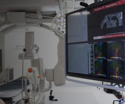
This layered approach enables AI-powered imaging technologies to build a detailed snapshot of vast volumes of data, layering images together to fill in gaps that are less defined due to patient movement, breathing, or coughing, which can create missing information or blurred pictures.
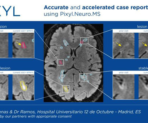
Pixyl is an award-winning French medtech specializing in AI-powered MRI solutions to improve patientcare. With disease modifying treatments for MS and now Alzheimer's Disease, it is more important than ever to highlight activity and monitor disease evolution.” For more information: www.pixyl.ai
christine.book Mon, 12/04/2023 - 11:29 December 4, 2023 — Ajay Gupta, MD, MS , has been named chair of the Department of Radiology at Columbia University Vagelos College of Physicians and Surgeons and radiologist-in-chief at NewYork-Presbyterian/CUIMC.
The rising demand for imaging services, coupled with an aging population and ever-increasing shortage of radiologists, creates the perfect storm for backlogs on a reading list. Clearing backlogs has become part and parcel of a diagnostic radiologists workflow. Even at six months, about 20% of these studies remained unreported.
This increase recognizes the growing importance of CCTA in diagnosing coronary artery disease and aligns reimbursement with the true cost of performing these procedures (American College of Radiology, 2024). Vesta provides access to subspecialized radiologists in cardiology, musculoskeletal imaging, neuroradiology, and more.
Benefits of Teleradiology to Telehealth Introduction: In the ever-evolving landscape of healthcare, smart imaging strategies are crucial for delivering improved patientcare. Explore the impact on patientcare when healthcare providers can tap into a network of expert radiologists for precise and timely interpretations.
Radiologists play a crucial role in healthcare, using advanced imaging technologies to diagnose and treat diseases. Their expertise is vital for maximizing patientcare by bridging the gap between clinical assessments and the detailed insights only imaging can provide.
milla1cf Tue, 06/18/2024 - 20:07 June 18, 2024 — The advancement of Artificial Intelligence ( AI ) in healthcare to support diagnostic decision making, stave off burnout and enhance quality patientcare has taken a giant stride forward. Samir Shah , Chief Medical Officer at Qure.ai Recently, Qure.ai
Introduction: Radiologists play a crucial role in modern healthcare by interpreting medical images to diagnose and guide patient treatment. To enhance diagnostic accuracy, reduce malpractice risk, and provide the highest level of patientcare, radiologists employ search patterns in their image interpretation processes.
Teleradiology & Radiology data for artificial intelligence (AI) Introduction: Nestled in the heart of the breathtaking Alps, Austria is revolutionizing patientcare through the adoption of teleradiology services. This multidisciplinary approach enhances patientcare, treatment planning, and overall healthcare outcomes.
February 28th is Rare Disease Day, an initiative created by the National Organization for Rare Disorders (NORD), to raise awareness and advocate for rare diseases. A rare disease is defined as a condition that affects fewer than 200,000 people in the United States, and there are over 7,000 rare diseases that have been identified.
Recent advancements in AI have revolutionized the way healthcare professionals approach AA care – both in the domain of thoracic aortic aneurysm (TAA) and abdominal aortic aneurysm (AAA) – leading to significant improvements in patient outcomes. However, many patients were lost to follow-up due to this cumbersome process.
A recent University of Washington School of Medicine study , presented at the 2024 Radiological Society of North America (RSNA) annual meeting, explored how integrating advanced AI technologies can help enhance radiologists’ performance. minutes, and attending radiologists times reduced from 6.6 Heres what they found.
Augmento X-Ray is designed to significantly reduce radiologist workload and improve the quality of chest X-ray reporting. Chest radiography is the most common medical imaging tool used in routine clinical practices to identify different disease findings. billion annual X-rays performed, 1.5
a provider of enterprise follow-up management and discovery software, announces the ability of their Discovery360 services to offload the follow-up responsibility from radiology and enable the entire health system to efficiently participate in the management of patients' Actionable Incidental Findings (AIFs).
This article delves into how teleradiology is enhancing patientcare across the vast Australian landscape, improving healthcare practices, and making diagnostic services accessible to all residents. This multidisciplinary approach enhances patientcare, treatment planning, and overall healthcare outcomes.
[i] ClearRead CT with CVI utilizes the latest advances in deep learning for a more predictive and preventative approach to patientcare. With ClearRead CT, radiologists can precisely detect, characterize, and report findings to improve diagnostic accuracy and advance earlier detection.”
The availability of these offerings – including the MIM SurePlan and MIM Symphony families, MIM Maestro, MIM Encore, and more – is in alignment with GE HealthCare’s precision care strategy, which aims to deliver innovative digital solutions across care pathways for more precise, connected, and efficient care across disease states. "We
Under Kontos’ leadership, the University reports that CIMBID will be dedicated to developing and integrating quantitative imaging and non-imaging biomarkers for disease prediction, particularly in cancer. and figures out a course of action.
We organize all of the trending information in your field so you don't have to. Join 5,000 users and stay up to date on the latest articles your peers are reading.
You know about us, now we want to get to know you!
Let's personalize your content
Let's get even more personalized
We recognize your account from another site in our network, please click 'Send Email' below to continue with verifying your account and setting a password.


Let's personalize your content