This site uses cookies to improve your experience. To help us insure we adhere to various privacy regulations, please select your country/region of residence. If you do not select a country, we will assume you are from the United States. Select your Cookie Settings or view our Privacy Policy and Terms of Use.
Cookie Settings
Cookies and similar technologies are used on this website for proper function of the website, for tracking performance analytics and for marketing purposes. We and some of our third-party providers may use cookie data for various purposes. Please review the cookie settings below and choose your preference.
Used for the proper function of the website
Used for monitoring website traffic and interactions
Cookie Settings
Cookies and similar technologies are used on this website for proper function of the website, for tracking performance analytics and for marketing purposes. We and some of our third-party providers may use cookie data for various purposes. Please review the cookie settings below and choose your preference.
Strictly Necessary: Used for the proper function of the website
Performance/Analytics: Used for monitoring website traffic and interactions
The Power of Diagnostic Imaging in Early Disease Detection Medical imaging is one of the most significant developments in medical science. By allowing doctors to view the inside of the body without the need for surgery, this technology aids in the early detection of diseases. How Does Early Detection Help?
Columbia University researchers in New York City have reported that worsening depression in older adults is related to higher levels of Alzheimers disease pathology, specifically the accumulation of tau protein. The study was published April 9 in the American Journal of Geriatric Psychiatry. The full study can be found here.
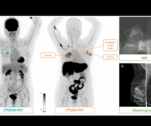
F-18 fluoroestradiol (FES)-PETscans can predict whether or not endocrine therapy may be effective in women with estrogen receptor (ER)-positive breast cancer, according to a study published March 13 in the Journal of Nuclear Medicine. A visual abstract of the analysis.
CHICAGO -- Characterizing an individual's type of body fat using body MRI can help predict Alzheimer's disease risk up to 20 years before symptoms manifest, according to research results presented December 2 at the RSNA meeting.
Preliminary studies suggest the tracer may also be effective in patients with earlier, more treatable stages of the disease, yet most of these studies have been retrospective and subject to a large risk of bias, the authors noted. The tumor in the right breast (T4N0M0, grade 3 ductal carcinoma) is visible on both PETscans.
Alzheimer’s disease patients with cognitive impairment or dementia who were referred for amyloid PETscans had fewer hospitalizations compared with a matched control group, according to a study published October 9 in JAMA Neurology. Each participant was matched to a control Medicare beneficiary who had not undergone amyloid PET.
Men should have prostate-specific membrane antigen (PSMA) PETscans about eight months after undergoing external beam radiotherapy for prostate cancer, a group at the University of California, Los Angeles has reported. The median duration between the first PETscan and the end-date of radiotherapy was 2.3
In a recent PET study with unexpected results, patients infected with herpes had fewer signs of brain deposits associated with Alzheimers disease than uninfected patients, a group in France has reported. The study was published January 18 in Scientific Reports. The median age of the group was 74 years old.
Patients with less tau pathology on PETscans may respond better to treatment with the new Alzheimer’s disease drug donanemab, according to an October 25 news report in the journal Practical Neurology. Donanemab is a monoclonal antibody designed to bind to beta-amyloid protein that already has formed into plaque deposits.
F-18 florbetaben PETscans for diagnosing Alzheimer’s disease can be reduced from 20 minutes to five minutes without losing accuracy, according to a recent study. Our findings suggest that shorter scan times are a viable and effective option for brain amyloid PET imaging in clinical settings,” the group wrote.
PETscans have revealed specific brain pathology that may drive sleep disturbances in Alzheimer’s disease patients, according to a study published February 8 in the Journal of the Neurological Sciences. Of these, 119 (43%) had normal cognition, 132 (24%) had mild cognitive impairment, and three (2%) had Alzheimer’s disease.
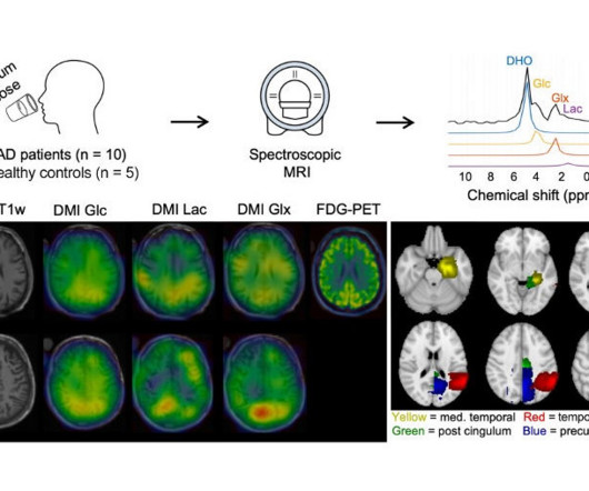
Deuterium metabolic imaging (DMI) was highly aligned with F-18 FDG-PET -- a cornerstone of dementia diagnostics -- in patients with Alzheimer's disease in a study published April 8 in Radiology. Given that PETscans are more expensive and expose patients to small amounts of radiation, DMI is being explored as an alternative.
Food and Drug Administration (FDA) has approved Eli Lilly's drug donanemab (Kisunla) for the treatment of early Alzheimer's disease, including mild cognitive impairment or mild dementia. According to clinical trials, amyloid PETscans showed that donanemab reduced amyloid plaques by up to 84% after 18 months of treatment.
PET imaging using a newly developed radiotracer has identified different patterns of brain tau pathology over time in early-onset versus late-onset Alzheimer’s disease patients, according to a study published February 1 in the Journal of Nuclear Medicine. in 15 patients with negative amyloid PETscans; 1.18 in
Kuo, who led development of imaging guidelines used to develop Lu-177 PSMA-617 ( Pluvicto , Novartis), noted that the theranostics treatment is approved for use in advanced stages of the disease, but that trials are underway to test it in patients at earlier stages. “We last year for Alzheimer's disease.
The agency is looking to lift the longstanding NCD that restricts patients to one amyloid-detecting positron emission tomography exam in their lifetime.
Read more on AuntMinnie.com Related Reading: FDA grants full approval to Alzheimer's disease drug Leqembi CMS rethinks limit on PETscans for Alzheimer’s disease patients FDA doubles MRI scans needed for Aduhelm patients CMS to limit coverage of new drugs for Alzheimer's disease CMS scrubs rule restricting PET tracer coverage
PET brain scans show persistent brain inflammation in patients with multiple sclerosis (MS), despite being treated with high-efficacy disease-modifying therapies, according to a recent study by researchers in Boston. The researchers performed F-18 PBR06 PETscans on 22 patients with MS and eight healthy controls.
Centers for Medicare and Medicaid Services (CMS) announced on October 13 that it has lifted its coverage limit of one beta-amyloid PETscan per lifetime for patients with Alzheimer’s disease. Medicare coverage decisions for amyloid PETscans will now be made by local Medicare Administrative Contractors (MACs).
Chinese clinicians have provided evidence in a “real-world study” that shows amyloid PET imaging is effective for diagnosing and managing patients with Alzheimer’s disease, according to a study published February 8 in Alzheimer’s and Dementia. of patients, noted lead author Ke-Liang Chen, MD, a neurologist at Fudan University. “In
A PET study has revealed that women over the age of 70 who took menopausal hormone therapy (HT) more than a decade before have faster brain tau accumulation -- a key indicator of Alzheimers disease. The findings may inform [Alzheimers disease] risk discussions relating to womens reproductive health and treatment, the group wrote.
In a small study, researchers at the National Institutes of Health have found that positron emission tomography (PET) scans of the heart may identify people who will go on to develop Parkinson's disease or Lewy body dementia among those at-risk for these diseases.
Early brain accumulation of amyloid plaque on PETscans is associated with emerging depressive symptoms in cognitively unimpaired older adults, according to a study published August 29 in JAMA Network Open. Their aim in this study was to determine whether these associations persist or progress over time.
A group in France has offered an explanation for long COVID brain fog, with the concept based on a visual brain pattern they discovered on patient PETscans, according to a study published October 13 in Medical Hypotheses. “We A) PETscan of a healthy subject. (B) B) PETscan of a long COVID patient.
The new radiotracer, called FDT, enables PETscans to be used for the first time to accurately pinpoint when and where the disease is still active in a patient's lungs. Radiotracers are radioactive compounds which give off radiation that can be detected by scanners and turned into a 3D image.
A PET radiotracer for diagnosing Alzheimer’s disease may also be used to measure vascular brain changes in patients during PET/MRI scans, according to a study published December 7 in the Journal of Nuclear Medicine.
Microglia are immune cells in the brain that are thought to have a role in MS disease progression but cannot be seen by a routine MRI. The team developed a technique called F18 PBR 06 PET imaging. The newly published study involved performing PETscans on 22 people with MS and eight healthy controls.
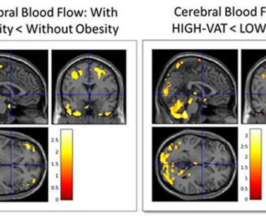
People with higher amounts of visceral abdominal fat in midlife may be at increased risk of Alzheimer's disease, according to research to be presented at the upcoming RSNA meeting. A subset of 32 patients underwent PET imaging to identify any amyloid plaque and tau tangles that would indicate the development of Alzheimer's disease.
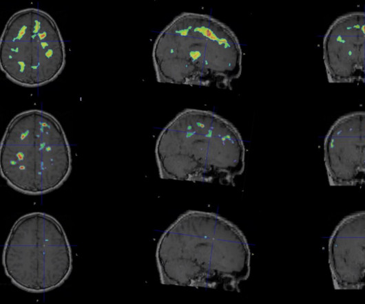
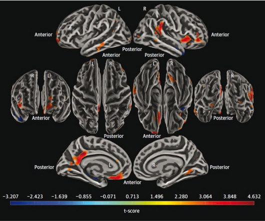
A PET/MRI study has provided insights into the neurobiology of late-life depression, with researchers reporting that tau protein – a key protein involved in Alzheimer’s disease – does not appear to be involved in the condition. Image and caption courtesy of the American Journal of Geriatric Psychiatry.
MRI is the imaging gold standard for diagnosis, yet identifying the disease using this method remains challenging, the researchers wrote. In all three cases, gadolinium-enhanced MRI scans did not show abnormalities. F-18 FDG-PET and MRI scans of three patients. In our outpatient clinic follow-up (2 to 2.5
PET imaging has revealed brain pathology linked to faster clinical progression of Alzheimer’s disease in patients with ApoE4 gene variants, according to a study published November 6 in JAMA Neurology.
In a proof-of-concept study, the researchers used F-18 FDG-PETscans in healthy participants within minutes after they performed standing and walking tasks and identified specific changes in brain glucose metabolism. F-18 FDG-PETscans allow clinicians to measure the brain's energy demands based on glucose metabolism.
PETscans typically used for diagnosing prostate cancer may have value in detecting inflammatory bowel disease (IBD), according to a case series published December 8 in Clinical and Experimental Gastroenterology.
Read more on AuntMinnie.com Related Reading: CMS to cover Alzheimer's drugs for patients enrolled in registry Will lecanemab approval increase PETscan volumes? FDA approves new drug for Alzheimer's disease CMS delays decision on amyloid PET coverage MRI sheds light on effects of aducanumab Alzheimer's drug
Participants underwent both F-18 PSMA-1007 PET/CT and multiparametric MRI within 14 days of one another and at least five days before surgery at two hospitals in Alberta, Canada. Out of 150 men who participated, 134 ultimately underwent radical prostatectomies (the mean age was 62 years old).
Read more on AuntMinnie.com Related Reading: CMS to cover Alzheimer's drugs for patients enrolled in registry Will lecanemab approval increase PETscan volumes? FDA approves new drug for Alzheimer's disease FDA doubles MRI scans needed for Aduhelm patients MRI sheds light on effects of aducanumab Alzheimer's drug
Read more on AuntMinnie.com Related Reading: FDA grants full approval to Alzheimer's disease drug Leqembi MITA chides CMS on beta-amyloid PET decision CMS to cover Alzheimer's drugs for patients enrolled in registry FDA approves new drug for Alzheimer's disease CMS delays decision on amyloid PET coverage
In this case, a 43-year-old man underwent an MRI scan that showed no contrast enhancement, yet hyperintensities were apparent in the patient’s left thalamus and frontoparietal region. Thus, the clinicians performed an additional PETscan with an amino acid radiotracer (F-18 FET) for further diagnosis of a suspected glioma.
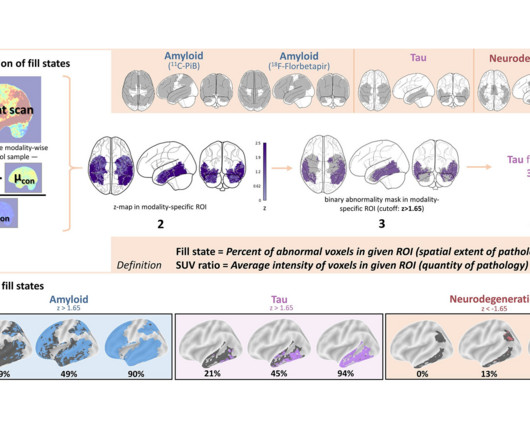
Researchers in Germany have proposed a new approach derived from brain PET imaging for diagnosing and staging Alzheimers disease, according to a study published March 25 in Radiology. They computed fill states for amyloid, tau, and neurodegeneration using percentages of significantly abnormal voxels, the smallest unit in a PET image.
The technology could allow clinicians to detect multiple biomarkers at once and improve the spatial resolution of brain imaging for applications in cancer as well as neurodegenerative disease, according to principal investigator Lars Furenlid, PhD, a professor of medical imaging at the University of Arizona.
Centers for Medicare and Medicaid Services (CMS) proposed a new reimbursement plan July 10 for diagnostic PETscans that would provide separate payments for radiopharmaceuticals, as well as an extra payment for hospitals when they use domestically produced technetium-99m (Tc-99m).
Read more on AuntMinnie.com Related Reading: NaF-PET shows bone formation in psoriatic arthritis patients PET/MRI provides new insights into knee osteoarthritis NaF-PET reveals aortic wall injuries NaF-PETscans reveal plaque -- and possible risk of stroke Can deep learning monitor lesions on F-18 NaF PET/CT?
We organize all of the trending information in your field so you don't have to. Join 5,000 users and stay up to date on the latest articles your peers are reading.
You know about us, now we want to get to know you!
Let's personalize your content
Let's get even more personalized
We recognize your account from another site in our network, please click 'Send Email' below to continue with verifying your account and setting a password.


Let's personalize your content