This site uses cookies to improve your experience. To help us insure we adhere to various privacy regulations, please select your country/region of residence. If you do not select a country, we will assume you are from the United States. Select your Cookie Settings or view our Privacy Policy and Terms of Use.
Cookie Settings
Cookies and similar technologies are used on this website for proper function of the website, for tracking performance analytics and for marketing purposes. We and some of our third-party providers may use cookie data for various purposes. Please review the cookie settings below and choose your preference.
Used for the proper function of the website
Used for monitoring website traffic and interactions
Cookie Settings
Cookies and similar technologies are used on this website for proper function of the website, for tracking performance analytics and for marketing purposes. We and some of our third-party providers may use cookie data for various purposes. Please review the cookie settings below and choose your preference.
Strictly Necessary: Used for the proper function of the website
Performance/Analytics: Used for monitoring website traffic and interactions
. | M7-SSPH05-2 | Room N229 Findings will be presented in this Monday afternoon presentation on organ-specific ionizing radiation doses in neonatal patients who undergo interventional procedures for congenital heart disease (CHD). Gy-cm2, with organ-specific radiation doses highest for lung from frontal view (8.1
Louis asked, “How much ionizing radiation are neonatal patients exposed to during interventional procedures?” Radiation doses estimated in infants with congenital heart disease Monday, November 27 | 3:10 p.m.-3:20 In one, a group at the University of Washington in St. 3:20 p.m. | 8:40 a.m. | 8:30 a.m. | 1:50 p.m. |
His research interests include using structural and functional MRI -- particularly ultrahigh-field, 7-tesla MRI -- to map brain microstructure and develop neurosurgical treatment of brain tumors, epilepsy, and neurodegenerative and movement disorders such as Parkinson's disease, essential tremor, and dystonia.
Chest dynamic digital radiography (DDR) may have received a boost toward clinical use in patients with lung disorders, with researchers developing AI to perform time-consuming analysis involved in the technology, according to researchers in New York City.
milla1cf Tue, 09/26/2023 - 15:32 September 26, 2023 — In a study of more than 2,000 chest X-rays , radiologists outperformed AI in accurately identifying the presence and absence of three common lung diseases, according to a study published in Radiology , a journal of the Radiological Society of North America ( RSNA ).
Led by Eli Atar, MD, director of the department of imaging, and Ahuva Grubstein, MD, department of diagnostic radiology, the study will assess the diagnostic capabilities of the Nanox.ARC’s tomographic imaging system compared with conventional 2D radiography for detecting lung and chest disease in adults.
Dynamic chest radiography (DCR) shows potential as a tool to investigate lung health in people with cystic fibrosis (CF), according to research published February 13 in Clinical Radiology. and colleagues.
X-ray radiography is a noninvasive diagnostic method that uses X-rays—electromagnetic radiation—to produce images of the body's internal structures. Essential in medical fields for diagnosing injuries and diseases and monitoring treatment progress, X-ray radiography is a critical tool in patient care and medical decision-making.
milla1cf Fri, 02/23/2024 - 10:22 February 23, 2024 — The American Society of Radiologic Technologists (ASRT) launched its "Be Seen" campaign today to raise public awareness about the crucial role medical imaging and radiation therapy professionals play in patient diagnosis, intervention and treatment.
For example, IEC Exposure Index allows quick assessment of the amount of radiation used to create the image; while Deviation Index immediately compares the chosen exposure to your facility’s specific target goal. The result: image quality comparable to images acquired with an anti-scatter grid but at a lower radiation dose.
Planning for Lung Disease Scanning Between late 2023 and early 2024, AIxSCAN, Inc. plans to produce over 50 lung disease patients scans in the U.S. and up to 1,000 total lung disease patients scans within 2 years. In addition, AIxSCAN, Inc. AIxSCAN, Inc. The company update specified that AIxSCAN, Inc.
This aids in the early detection and monitoring of diseases. Hybrid Imaging : The integration of multiple imaging modalities, such as PET/CT and SPECT/CT, allows for more accurate diagnosis and staging of diseases, particularly in oncology.
Not only are children more radiosensitive than adults (the cancer risk per unit dose of ionizing radiation is higher), but children also have a longer expected lifetime, which puts them at greater risk of cancer following radiation exposure.(1) The balance of dose and image quality is even more important in pediatric medical imaging.
Chapter 3: The Radiologic Toolbox – Types of X-ray Imaging An exploration of the various types of X-ray imaging, including radiography, fluoroscopy, computed tomography (CT), and more. The importance of minimizing radiation exposure while maintaining diagnostic accuracy.
The principles of radiation and how X-rays interact with the human body to create diagnostic images. Chapter 3: Types of X-ray Imaging: Beyond Radiography An exploration of the various types of X-ray imaging, including radiography, fluoroscopy, and computed tomography (CT).
Radiation oncology clinics face numerous challenges in the present environment, including the simultaneous management of multiple tasks (many of which are manual in nature), various degrees of standardisation, and the potential for errors to impact patient treatment. per week for curative diseases.
Additionally, the organization reported it launched its ASRT “Be Seen” public awareness campaign in late February to raise awareness about the crucial role medical imaging and radiation therapy professionals play in patient diagnosis, intervention and treatment. ASRT 2024-2025 Board of Directors Daniel DeMaio, M.Ed., Jennifer Thompson, Ed.D.,
Digital Transformation : Traditional film-based radiography is being replaced by digital imaging systems, which offer higher resolution, more efficient data management, and the ability to share images electronically.
Traditional film-based X-rays gave way to digital radiography (DR) and computed radiography (CR). They are used in the diagnosis of bone fractures, lung diseases, dental issues, and various other medical conditions. Advanced Imaging Modalities: X-ray technology has expanded beyond conventional radiography.
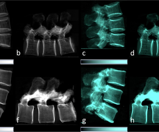
“This finding implies the potential application of dark-field imaging to draw conclusions on bone microstructure for predicting bone stability at a lower radiation exposure than in tomographic modalities,” wrote Jon Rischewski, a doctoral candidate at Ludwig Maximilian University of Munich, and colleagues.
Ultrasound model predicts liver disease progression. Commercially Available Chest Radiograph AI Tools for Detecting Airspace Disease, Pneumothorax, and Pleural Effusion. Effects of low-dose ionizing radiation on genomic instability in interventional radiology workers. Shah, et al, Radiography , January 9, 2024.
We organize all of the trending information in your field so you don't have to. Join 5,000 users and stay up to date on the latest articles your peers are reading.
You know about us, now we want to get to know you!
Let's personalize your content
Let's get even more personalized
We recognize your account from another site in our network, please click 'Send Email' below to continue with verifying your account and setting a password.


Let's personalize your content