This site uses cookies to improve your experience. To help us insure we adhere to various privacy regulations, please select your country/region of residence. If you do not select a country, we will assume you are from the United States. Select your Cookie Settings or view our Privacy Policy and Terms of Use.
Cookie Settings
Cookies and similar technologies are used on this website for proper function of the website, for tracking performance analytics and for marketing purposes. We and some of our third-party providers may use cookie data for various purposes. Please review the cookie settings below and choose your preference.
Used for the proper function of the website
Used for monitoring website traffic and interactions
Cookie Settings
Cookies and similar technologies are used on this website for proper function of the website, for tracking performance analytics and for marketing purposes. We and some of our third-party providers may use cookie data for various purposes. Please review the cookie settings below and choose your preference.
Strictly Necessary: Used for the proper function of the website
Performance/Analytics: Used for monitoring website traffic and interactions
Most radiologists have heard of Moore’s Law. A close colleague observed that radiologists are affected by a type of Moore’s Law. The amount of scans a radiologist is expected to shift per day is increasing year on year. Today’s radiologist is expected to shift work at an eye-watering rate. Paul McCoubrie, MBBS.
The voters also zeroed in on the ongoing shortage of radiologists as the Biggest Threat to Radiology. Importantly, we are gaining deeper insights into disease processes themselves, which enhances our ability to diagnose and potentially treat conditions that were previously beyond our reach." I'm a radiographer,' " Stewart recalled.
Understanding the mechanics of flow artifacts on CT or CT angiography (CTA) and how these artifacts are created is key to better disease diagnosis, according to a review published April 25 in RadioGraphics. In the review, a team led by Caroline Robb, MD, of Mallinckrodt Institute of Radiology, Washington University in St.
AI for thoracic imaging includes using it for reading chest radiographs and low-dose chest CT scans for lung cancer screening and for triaging pulmonary embolism on chest CT scans, the group noted. Next-generation AI technology shows promise, however.
Among questions that remain unanswered about AI is whether the quality of its mistakes is different than those of radiologists and if AI mistakes, on average, are objectively worse than human mistakes. These included chest radiographs that displayed abnormalities of no clinical significance, which are typically treated as normal.
AI methods improve detection of Parkinson’s disease Sunday, November 26 | 1:50 p.m.-2:00 S4-SSNR02-6 | Room E352 A systematic review has found that machine-learning and deep-learning techniques are highly sensitive and specific for detecting Parkinson’s disease on dopamine transporter (DaT) SPECT exams. 2:00 p.m. | 1:40 p.m. |
Healthcare disparities continue to plague medical imaging, but there are concrete measures radiologists can take to mitigate them, according to a paper published on October 12 in RadioGraphics. Histopathologic biopsy results demonstrated invasive mammary carcinoma with metastatic disease in a right axillary lymph node.
Imaging was so important [for cardiac indications], that I decided to become a radiologist," he said. Together with our radiographers, I learned to scan cardiac patients and learned special anatomy from pediatric cardiologists and pediatric cardiac surgeons." How can radiologists become successful cardiac imagers? Just do it!"
A team led by Junqi Han, MD, from the Affiliated Hospital of Qingdao University in China found that its model combining data from mammography images, ultrasound images, and other characteristics performed well in predicting disease-free survival of breast cancer.
Alzheimer disease is a progressive, irreversible brain disorder that slowly degrades memory and cognitive function. While previous treatment methods focused on addressing Alzheimer disease symptoms, recent approvals of monoclonal antibodies have provided a path to target the underlying disease itself. In June 2021, the U.S.
S1-SSCH01-2 | E451A Computing thoracic bone and muscle metrics in patients with chronic obstructive pulmonary disease (COPD) improves understanding of the role of certain comorbidities in COPD disease progression, according to findings to be announced in this scientific presentation. 9:20 a.m. | 10:50 a.m. | 11:00 a.m. | 2:40 p.m. |
The American Roentgen Ray Society (ARRS) is recognizing four radiologists, as well as their institutions and research projects, with its 2024 ARRS Resident/Fellow in Radiology Awards. The following radiologists and their research projects are recognized as awardees: Melina Hosseiny, MD, University of California, San Diego.
i Thankfully, the field of breast screening is not static, and advancements are revolutionizing how we detect and diagnose the disease. New AI integrations are further elevating the impact of this technology, not only by acting as a valuable second set of eyes for radiologists but also by providing standardized breast density assessments.
His research interests include using structural and functional MRI -- particularly ultrahigh-field, 7-tesla MRI -- to map brain microstructure and develop neurosurgical treatment of brain tumors, epilepsy, and neurodegenerative and movement disorders such as Parkinson's disease, essential tremor, and dystonia.
The rising demand for imaging services, coupled with an aging population and ever-increasing shortage of radiologists, creates the perfect storm for backlogs on a reading list. At seven days post-imaging, nearly half had unreported brain and chest CT scans, while 59% had unreported chest radiographs. The consequences are significant.
The Society publishes five established journals: Radiology, RadioGraphics, Radiology: Artificial Intelligence, Radiology: Cardiothoracic Imaging and Radiology: Imaging Cancer. RadioGraphics , RSNA’s premier educational journal, edited by Christine ‘Cooky’ Menias , M.D., Klein , M.D., RSNA Board Liaison for Publications.
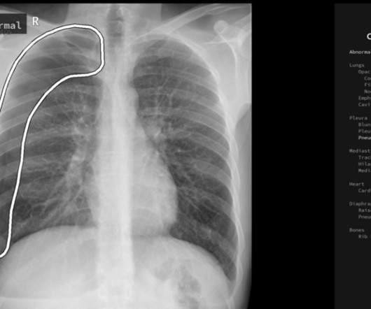
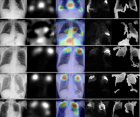
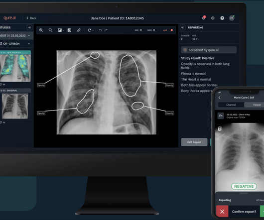
Augmento X-Ray is designed to significantly reduce radiologist workload and improve the quality of chest X-ray reporting. Chest radiography is the most common medical imaging tool used in routine clinical practices to identify different disease findings. billion annual X-rays performed, 1.5
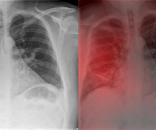
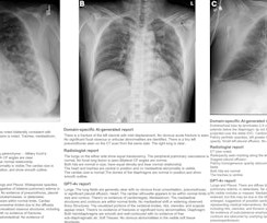
However, the sensitivity of a chest radiograph in the diagnosis of COVID-19 is moderate. Therefore, in this study, the authors compared the performance of a deep learning algorithm (Ensemble4Covid) on the first clinical encounter with a group of radiologists with varying years of experience. González Montaño, Clara E.
Cooky’s tough love: It takes time to become an excellent radiologist. We should be leveraging the ‘free’ body composition data that is available on all abdominal CTs.”— Learn from your mistakes and the mistakes of others. Your practice will change when it becomes personal.”—Christine
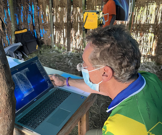
Rob Liddell, MD, is a diagnostic radiologist who used MinXray’s Impact Wireless system to take radiographic images of patients in the village and screen for common diseases in the region, such as tuberculosis and emphysema. He also used the system to diagnose cancers, infections and various musculoskeletal injuries.
A fundamental goal of radiographers is to complete an imaging exam that provides sufficient information for an accurate clinical diagnosis–and at the lowest possible dose. We also have a Detector Verification alert that signals radiographers when they choose the wrong detector–which would result in the need to repeat the exam.
But how will AI in the workplace affect the radiographer and how does it differ from the red dot system radiographers are so familiar with? The Red Dot System Often one of the first courses a newly qualified radiographer attends is the red dot course. This is used to create an alert for the referring clinician/radiologist.
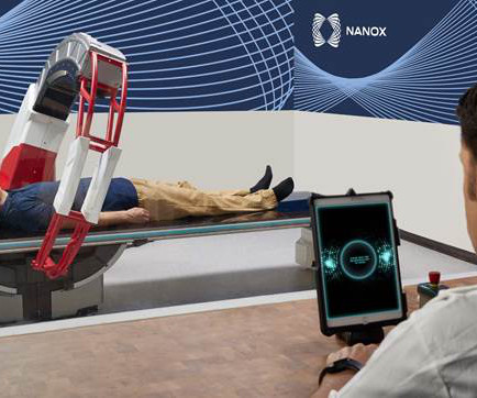
Medical imaging is essential for detecting, diagnosing, and managing disease, guiding treatment decisions for improved health outcomes. Following this clearance, Nanox will continue to work with the FDA to pursue additional regulatory clearances and intends to expand clinical indications.
The approval expands upon Bayer's focus on breast imaging, with a portfolio that also includes Gadavist (gadobutrol) injection, a gadolinium-based contrast agent approved for use with MRI ( Magnetic Resonance Imaging ) to assess the presence and extent of malignant breast disease in adult patients. RadioGraphics 2019 39:7, 1907-1920.
Radiology is a branch of medicine that uses radiant energy in the diagnosis and treatment of disease. Practitioners of radiology are called radiologists, and they utilize imaging technology in the diagnosis and treatment of patients. Medical imaging is a technology which is used by radiologists , particularly for diagnostic purposes.
The Essentia SA is an ultra-compact straight arm system, designed for a wide range of standing, sitting and recumbent radiographic exams. Both systems feature Fujifilm’s dose performance technologies to capture superior images at a low dose for the patient providing exception price to performance ratio.
Changes in the standard of care for cardiac imaging means there’s an increasing need for radiologists to be able to read cardiac CT and MRI. Free Webinar: 1 Heart. AMA PRA Category 1 Credits Focus Your Training with our Cardiac MRI and CT Library Plan!
A B Reader is a physician who has been certified by the National Institute for Occupational Safety and Health (NIOSH) for showing expertise in classifying radiographs related to pneumoconioses. Their primary role is to identify potential occupational-related diseases in patients.
A) Dorsoplantar radiograph of the foot demonstrating an isolated fracture of the cuboid with possible extension into the tarsometatarsal joint. (B) B) Medial oblique radiograph of the foot demonstrating an isolated fracture of the cuboid. Radiographic evidence can support the diagnosis. Trauma in a 8 year old female.
Machine learning in healthcare represents a revolutionary approach to analyzing vast datasets to uncover patterns, predict disease states, flag suspected pathologies and personalize treatment plans. This capability leads to earlier disease awareness, enabling timely treatment and, ideally, improved patient outcomes.
This aids in the early detection and monitoring of diseases. Hybrid Imaging : The integration of multiple imaging modalities, such as PET/CT and SPECT/CT, allows for more accurate diagnosis and staging of diseases, particularly in oncology.
My name is Dr. Lori Barr and I am a practicing pediatric radiologist with Radiology Associates of Florida. We recently adopted a nationwide pediatric appendix ultrasound performance protocol, sonographer worksheet, and radiologist reporting template in order to decrease CT utilization for this diagnosis nationwide.
Frontal abdomen radiograph demonstrates foreign body consistent with capsule endoscopy device (pill cam) in descending colon. Capsule Retention and Risk Factors: Capsule endoscopy is used for evaluating small-bowel disorders, such as bleeds and Crohn disease.[ What are the important findings in this case. World J Gastroenterol.
Following surgery, patients should begin antibiotic coverage of pathogens associated with endophthalmitis per recommendations provided by their institution’s infectious disease specialist. Radiographics. Rice, MD is the president of Global Radiology CME and is a radiologist with Cape Radiology Group. Clin Ophthalmol.
This chapter deserves extra attention by the reader since there are pitfalls which can send the clinicians, surgeons, and radiologists to the wrong diagnosis. While these items are not usually thought of by radiologists, it is worthwhile to know what spine surgeons either now use or will be using in these difficult operations.
A team led by Eun Kyoung Hong, MD, PhD, from Brigham & Womens Hospital in Boston, MA, found that their domain-specific model could detect conditions such as pneumothorax and subcutaneous emphysema and achieved a high rate of reports accepted without modification from radiologists.
Randall Munroe, author of the always wonderful XKCD and Thing Explainer , illustrates why basically every radiologist should be doing this : Please note that I am far from an expert on AHK or scripting in general. insert normal chest radiograph), a button can be even more frictionless. (I
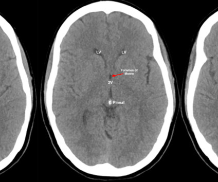
Written by: Justin Seltzer, MD (NUEM PGY-1) Edited by: Andrew Cunningham, MD, (NUEM PGY-3) Expert commentary by : David Rusinak, MD Neuroimaging, mainly using CT, has become an indispensable part of our emergency diagnostic process, but, all too often we rely on radiologists to interpret what we ordered. RadioGraphics, 1998; 18(1):151-163 3.
Centers for Disease Control and Prevention (CDC) is considering the use of AI to back up its tuberculosis (TB) screening program for immigrants and refugees. After COVID-19, TB is the second leading cause of death from infectious disease in the world. has relatively low rates – about 2.5 has relatively low rates – about 2.5 to 0.99).
Ultrasound model predicts liver disease progression. Commercially Available Chest Radiograph AI Tools for Detecting Airspace Disease, Pneumothorax, and Pleural Effusion. Generative Artificial Intelligence for Chest Radiograph Interpretation in the Emergency Department. SPECT/CT reveals heart’s response to tafamidis.
accurate labeling x-ray reports for thoracic diseases, but performed poorly in patients over 80 years old, noted lead author Samantha Santomartino, a medical student at Drexel University in Philadelphia, and colleagues. Three board-certified radiologists annotated the images for 14 thoracic disease labels.
The data will be available through the OSIC Data Repository to AI experts and other collaborators to design algorithms that could potentially identify novel biomarkers and relate radiograph quantifications to clinical indicators, and to disease risk and prognostication factors. 1) There is no known cause and no known cure.
Notably, this is the only FDA-cleared solution for detecting and localizing lung nodules utilizing computer vision to have Radiologists, Pulmonologists and ER physicians as intended users. The device's performance was assessed against the ground truth determined by five American board-certified Radiologists.
I believe that the first main difference seen in the radiologists role in 2040 will be the change that artificial intelligence (AI) will have made on the diagnostic role that radiologists currently perform. There are several protective factors, the first being that radiologists do much more than just report scans.
We organize all of the trending information in your field so you don't have to. Join 5,000 users and stay up to date on the latest articles your peers are reading.
You know about us, now we want to get to know you!
Let's personalize your content
Let's get even more personalized
We recognize your account from another site in our network, please click 'Send Email' below to continue with verifying your account and setting a password.


Let's personalize your content