This site uses cookies to improve your experience. To help us insure we adhere to various privacy regulations, please select your country/region of residence. If you do not select a country, we will assume you are from the United States. Select your Cookie Settings or view our Privacy Policy and Terms of Use.
Cookie Settings
Cookies and similar technologies are used on this website for proper function of the website, for tracking performance analytics and for marketing purposes. We and some of our third-party providers may use cookie data for various purposes. Please review the cookie settings below and choose your preference.
Used for the proper function of the website
Used for monitoring website traffic and interactions
Cookie Settings
Cookies and similar technologies are used on this website for proper function of the website, for tracking performance analytics and for marketing purposes. We and some of our third-party providers may use cookie data for various purposes. Please review the cookie settings below and choose your preference.
Strictly Necessary: Used for the proper function of the website
Performance/Analytics: Used for monitoring website traffic and interactions
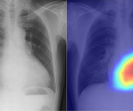
ChatGPT-4 outperformed human clinicians in determining pretest and post-test disease probability after a negative test result involving chest radiographs and mammograms, according to a research letter published December 11 in JAMA Network Open.
. | M7-SSPH05-2 | Room N229 Findings will be presented in this Monday afternoon presentation on organ-specific ionizing radiation doses in neonatal patients who undergo interventional procedures for congenital heart disease (CHD). Louis, and colleagues.
Exciting presentations will cover a generative AI model that shows potential for making surgical planning for total hip arthroplasty (THA) more efficient and a deep-learning model developed to identify individuals at high risk of chronic obstructive pulmonary disease (COPD). 3:20 p.m. | 8:40 a.m. | 8:30 a.m. | 1:50 p.m. |
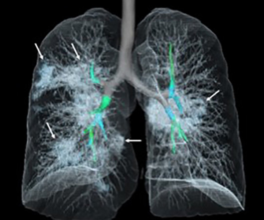
Researchers at the Icahn School of Medicine at Mount Sinai ("Icahn Mount Sinai") used Dynamic Digital Radiography (DDR) data, an X-ray imaging technology developed by Konica Minolta, to create their AI-powered technique that analyzes lung function. Chest radiography is typically acquired in the evaluation of pulmonary disorders.
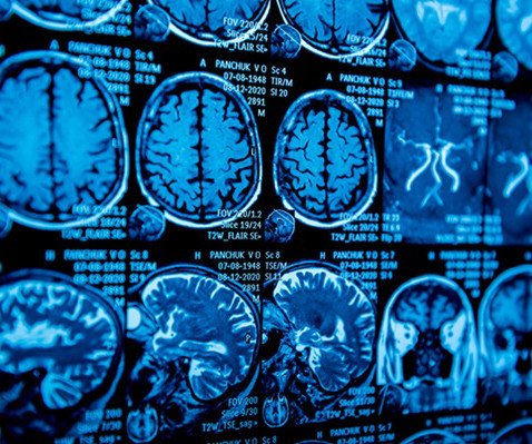
His research interests include using structural and functional MRI -- particularly ultrahigh-field, 7-tesla MRI -- to map brain microstructure and develop neurosurgical treatment of brain tumors, epilepsy, and neurodegenerative and movement disorders such as Parkinson's disease, essential tremor, and dystonia.
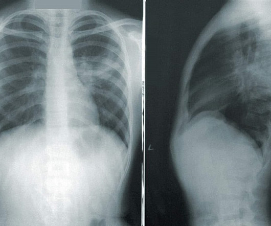
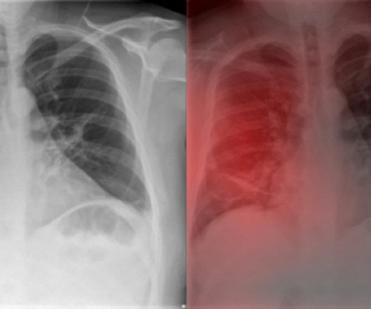
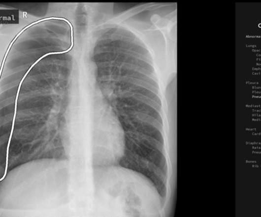
milla1cf Tue, 09/26/2023 - 15:32 September 26, 2023 — In a study of more than 2,000 chest X-rays , radiologists outperformed AI in accurately identifying the presence and absence of three common lung diseases, according to a study published in Radiology , a journal of the Radiological Society of North America ( RSNA ).
As technology continues to advance, physicians have a multitude of methods to diagnose diseases. Radiography is a broad field that uses imaging techniques to view the skin. Doctors use diagnostic radiography to non-invasively diagnose injuries and diseases inside the body. Enter diagnostic radiology.
Chest dynamic digital radiography (DDR) may have received a boost toward clinical use in patients with lung disorders, with researchers developing AI to perform time-consuming analysis involved in the technology, according to researchers in New York City.
Dynamic digital radiography (DDR) has shown for the first time that it can be used to automatically capture lung signal changes during forced breathing in patients with chronic obstructive pulmonary disease (COPD), according to a recent study. and Japanese developers wrote.
Read more on AuntMinnie.com Related Reading: Dynamic chest radiography reveals treatment response in CF patients Thirona launches AI algorithm for cystic fibrosis Ultrasound identifies kids at risk of CF liver disease Novel MRI scans could aid in cystic fibrosis CT can replace x-ray for pediatric cystic fibrosis patients
A new study has revealed that MRI scans depicting the structural and functional organization of the brain can predict the progression of brain atrophy in patients with early-stage, mild Parkinson's disease.
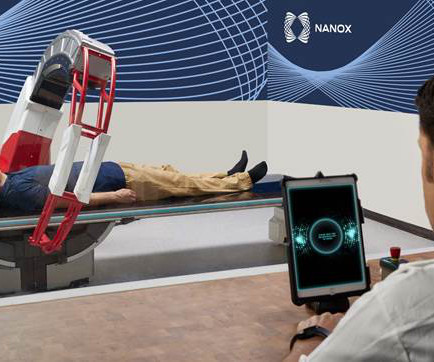
Led by Eli Atar, MD, director of the department of imaging, and Ahuva Grubstein, MD, department of diagnostic radiology, the study will assess the diagnostic capabilities of the Nanox.ARC’s tomographic imaging system compared with conventional 2D radiography for detecting lung and chest disease in adults.
announced significant expansion of X-Ray systems with Dynamic Digital Radiography (DDR) at multiple healthcare institutions across the US, including at Appleton Area Health (Appleton, Minn.), milla1cf Tue, 07/11/2023 - 08:37 July 11, 2023 — Konica Minolta Healthcare Americas, Inc., New Hampshire Neurospine Institute (Bedford, N.H.),
An innovative artificial intelligence tool uses chest X-rays to classify cardiac functions and identify valvular heart disease with unprecedented accuracy.
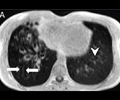
Dynamic chest radiography (DCR) shows potential as a tool to investigate lung health in people with cystic fibrosis (CF), according to research published February 13 in Clinical Radiology. and colleagues.
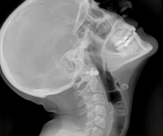
X-ray radiography is a noninvasive diagnostic method that uses X-rays—electromagnetic radiation—to produce images of the body's internal structures. Essential in medical fields for diagnosing injuries and diseases and monitoring treatment progress, X-ray radiography is a critical tool in patient care and medical decision-making.
Researchers have defined a set of ultrasound parameters that quantitatively evaluate various physical lung characteristics for a more accurate assessment.
Helps Doctors Monitor Disease Progression Diagnostic imaging is one of the most important markers that doctors utilize to monitor the progression of diseases for a variety of ailments, including: Certain types of malignant cancer Pneumonia and other respiratory diseases Internal bleeding Brain injuries Cardiac conditions Acute injuries (i.e.,
plans to produce over 50 lung disease patients scans in the U.S. and up to 1,000 total lung disease patients scans within 2 years. UC San Diego Health is the first site for the clinical trials to be conducted. Between late 2023 and early 2024, AIxSCAN, Inc. AIxSCAN, Inc. AIxSCAN, Inc.
A new computer-based diagnostic tool combines ultrasound imagery with specific clinical indicators to evaluate the risk of moderate-to-severe renal fibrosis.
The research will produce technical feedback to assist GEHC in assessing the system’s reconstruction methods, image presentation workflow, and clinical benefits for specific pathologies and disease types. It supports both radiography and fluoroscopic imaging. MRI GEHC unveiled Signa Champion, a 1.5-tesla,
The new imaging systems that will be on display include three new digital radiography (DR) suites, two new fluoroscopy systems, a 0.4T MRI system and a 128- slice computed tomography (CT) system. “At The Essentia SA is an ultra-compact straight arm system, designed for a wide range of standing, sitting and recumbent radiographic exams.
"While the tool cannot replace [DEXA] for osteoporosis screening, it can be a valuable option when lumbar spine radiography is readily available, and [DEXA] has not been performed," the authors wrote. Therefore, the clinical implementation of such a tool could enhance disease recognition and aid in preventing osteoporotic fractures."
Currently, no imaging procedure allows for therapy-relevant differentiation between intestinal wall inflammation and fibrosis. Now, a new imaging technique may.
Digital radiography utilizes a unique technology, similar to a digital camera, that instantly sends X-ray images to a computer for quick viewing. Likewise, digital radiography systems use a detector known as a flat panel to translate data into a digital electronic signal. This digital electronic signal becomes a digital X-ray image.
Chest radiography is the most common medical imaging tool used in routine clinical practices to identify different disease findings. Augmento X-Ray is designed to significantly reduce radiologist workload and improve the quality of chest X-ray reporting. billion annual X-rays performed, 1.5
An AI algorithm was presented that could make dynamic digital radiography (DDR) more efficient by automatically measuring kinematics involved in certain shoulder injuries. Research from a group in Germany was presented suggesting that an AI algorithm for pneumothorax detection can perform similarly to radiology resident readers.
A new study has shown that human radiologists were generally more accurate than AI in identifying the presence and absence of common lung diseases on chest X-rays.
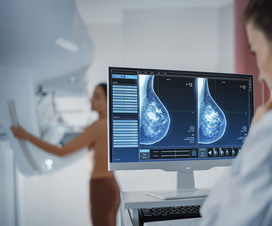
New studies have validated the ability of an AI-powered solution to uncover hidden heart diseases as well as predict a woman’s risk for developing breast cancer in the next one- or two years from a single mammogram.
Nanox.ARC is a stationary X-ray system intended to produce tomographic images of the human musculoskeletal system adjunctive to conventional radiography on adult patients. Medical imaging is essential for detecting, diagnosing, and managing disease, guiding treatment decisions for improved health outcomes.
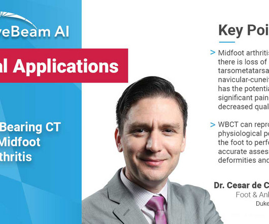
The most common etiology is posttraumatic degeneration, but primary degeneration and inflammatory diseases can also result in osteoarthritis of the midfoot joints. Comparative assessment of midfoot osteoarthritis diagnostic sensitivity using weightbearing computed tomography vs weightbearing plain radiography. Eur J Radiol.
UNC-Chapel Hill developed collaborative agreements with UNC Health and Duke Health for the planning and execution of two programs, a certificate program in radiography, and a bachelor of science degree program in diagnostic medical sonography. We should be leveraging the ‘free’ body composition data that is available on all abdominal CTs.”—
Planning for Lung Disease Scanning Between late 2023 and early 2024, AIxSCAN, Inc. plans to produce over 50 lung disease patients scans in the U.S. and up to 1,000 total lung disease patients scans within 2 years. In addition, AIxSCAN, Inc. AIxSCAN, Inc. The company update specified that AIxSCAN, Inc.
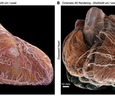
For the first time, researchers have used a synchrotron X-ray imaging technique to visualize two entire human adult hearts, both healthy and diseased, at the cellular level in 3D, uncovering previously unseen structures and connections.
Digital Tomosynthesis: a 3D extension of general radiography Digital Tomosynthesis (DT) is a three-dimensional extension of general radiography. References: 1 Unified Database for Rejected Image Analysis Across Multiple Vendors in Radiography. 2 During customer site evaluations using Carestream DR Smart Room Assist features.
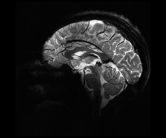
The world's most powerful MRI scanner has generated its first images of the human brain, demonstrating new precision levels that could shed more light on neurological diseases.
This was especially seen in the role of chest radiography when it was utilized as a diagnostic tool at the beginning of the pandemic “when microbiological resources were scarce,” evolving into its use focusing on the detection and monitoring of COVID-19 lung involvement.
This aids in the early detection and monitoring of diseases. Hybrid Imaging : The integration of multiple imaging modalities, such as PET/CT and SPECT/CT, allows for more accurate diagnosis and staging of diseases, particularly in oncology.
Chapter 3: The Radiologic Toolbox – Types of X-ray Imaging An exploration of the various types of X-ray imaging, including radiography, fluoroscopy, computed tomography (CT), and more. How X-rays are generated, interact with human tissue, and create diagnostic images. How each modality serves unique clinical purposes and applications.
Traditional film-based X-rays gave way to digital radiography (DR) and computed radiography (CR). They are used in the diagnosis of bone fractures, lung diseases, dental issues, and various other medical conditions. Advanced Imaging Modalities: X-ray technology has expanded beyond conventional radiography.
Chapter 3: Types of X-ray Imaging: Beyond Radiography An exploration of the various types of X-ray imaging, including radiography, fluoroscopy, and computed tomography (CT). Chapter 7: Evolving Technologies: From Analog to Digital The evolution of X-ray imaging technology, from analog film to digital radiography.
S hamie Kumar describes her perspective on how radiography has evolved over time, the impact radiographers can have in detecting abnormal X-rays and reflects how she views fast approaching AI in advancing current skills. The Red Dot System Often one of the first courses a newly qualified radiographer attends is the red dot course.
We organize all of the trending information in your field so you don't have to. Join 5,000 users and stay up to date on the latest articles your peers are reading.
You know about us, now we want to get to know you!
Let's personalize your content
Let's get even more personalized
We recognize your account from another site in our network, please click 'Send Email' below to continue with verifying your account and setting a password.


Let's personalize your content