This site uses cookies to improve your experience. To help us insure we adhere to various privacy regulations, please select your country/region of residence. If you do not select a country, we will assume you are from the United States. Select your Cookie Settings or view our Privacy Policy and Terms of Use.
Cookie Settings
Cookies and similar technologies are used on this website for proper function of the website, for tracking performance analytics and for marketing purposes. We and some of our third-party providers may use cookie data for various purposes. Please review the cookie settings below and choose your preference.
Used for the proper function of the website
Used for monitoring website traffic and interactions
Cookie Settings
Cookies and similar technologies are used on this website for proper function of the website, for tracking performance analytics and for marketing purposes. We and some of our third-party providers may use cookie data for various purposes. Please review the cookie settings below and choose your preference.
Strictly Necessary: Used for the proper function of the website
Performance/Analytics: Used for monitoring website traffic and interactions
This year’s trip along the Road to RSNA for digital x-ray features mileposts mostly set by AI research. Models will be proposed for applications ranging from predicting bone density on chest x-rays to generating complete reports on anterior cruciate ligament (ACL) tears. Nonetheless, AI is poised to take top headlines.
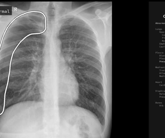
milla1cf Tue, 09/26/2023 - 15:32 September 26, 2023 — In a study of more than 2,000 chest X-rays , radiologists outperformed AI in accurately identifying the presence and absence of three common lung diseases, according to a study published in Radiology , a journal of the Radiological Society of North America ( RSNA ).
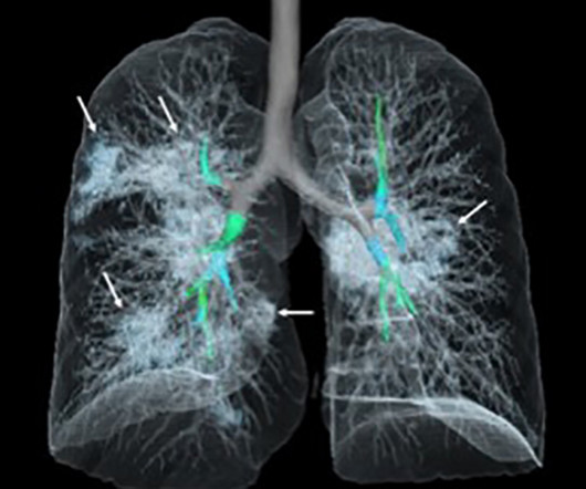
Chest dynamic digital radiography (DDR) may have received a boost toward clinical use in patients with lung disorders, with researchers developing AI to perform time-consuming analysis involved in the technology, according to researchers in New York City. The study was published March 29 in Chest Pulmonary.
Dear Digital X-Ray Insider, RSNA 2023 wrapped up in Chicago recently, with a bevy of digital x-ray studies presented at the meeting. We’ve featured one more here: an AI study conducted in Zambia testing an algorithm developed by Google to detect tuberculosis on chest x-rays. That's all for now!
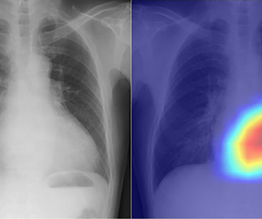
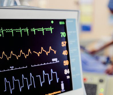
An innovative artificial intelligence tool uses chest X-rays to classify cardiac functions and identify valvular heart disease with unprecedented accuracy.
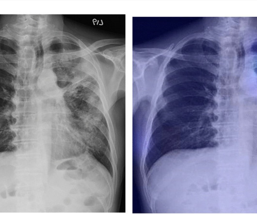
Dynamic digital radiography (DDR) has shown for the first time that it can be used to automatically capture lung signal changes during forced breathing in patients with chronic obstructive pulmonary disease (COPD), according to a recent study. and Japanese developers wrote. Dynamic image data was acquired at 7.5
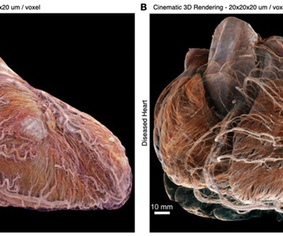
For the first time, researchers have used a synchrotron X-ray imaging technique to visualize two entire human adult hearts, both healthy and diseased, at the cellular level in 3D, uncovering previously unseen structures and connections.
. | M7-SSPH05-2 | Room N229 Findings will be presented in this Monday afternoon presentation on organ-specific ionizing radiation doses in neonatal patients who undergo interventional procedures for congenital heart disease (CHD). Louis, and colleagues.
Augmento X-Ray is designed to significantly reduce radiologist workload and improve the quality of chest X-ray reporting. Chest radiography is the most common medical imaging tool used in routine clinical practices to identify different disease findings. billion annual X-rays performed, 1.5
milla1cf Thu, 06/06/2024 - 21:32 June 6, 2024 — In a landmark study, the latest in technology innovation by Konica Minolta Healthcare was used to develop a machine-learning-based analysis of X-ray imaging that automatically quantifies lung function data. Chest radiography is typically acquired in the evaluation of pulmonary disorders.
Read more on AuntMinnie.com Related Reading: Dynamic chest radiography reveals treatment response in CF patients Thirona launches AI algorithm for cystic fibrosis Ultrasound identifies kids at risk of CF liver disease Novel MRI scans could aid in cystic fibrosis CT can replace x-ray for pediatric cystic fibrosis patients
How would you rate the value you are getting from your current X-ray imaging equipment? Upgrade your X-ray imaging equipment to get access to the most advanced imaging features for about 10% of the cost of original equipment. Does it support new medical imaging software that can help improve clinical outcomes?
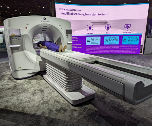
The scanner also comes with True Enhance DL, an AI-based application that generates deep learning-based monochromatic-like images from a single-energy x-ray acquisition. It supports both radiography and fluoroscopic imaging. GE HealthCare's new Revolution Ascend CT scanner. MRI GEHC unveiled Signa Champion, a 1.5-tesla,
X-rayradiography is a noninvasive diagnostic method that uses X-rays—electromagnetic radiation—to produce images of the body's internal structures. This technique allows healthcare professionals to examine bones, tissues, and organs without surgical intervention.
Dynamic chest radiography (DCR) shows potential as a tool to investigate lung health in people with cystic fibrosis (CF), according to research published February 13 in Clinical Radiology. and colleagues. Yet FEV1 may not always reflect the severity of the airway obstruction, they noted.
announced significant expansion of X-Ray systems with Dynamic Digital Radiography (DDR) at multiple healthcare institutions across the US, including at Appleton Area Health (Appleton, Minn.), A powerful X-ray generator and Automatic Exposure Control further optimize image quality and help minimize patient dose.
A new study has shown that human radiologists were generally more accurate than AI in identifying the presence and absence of common lung diseases on chest X-rays.
plans to produce over 50 lung disease patients scans in the U.S. and up to 1,000 total lung disease patients scans within 2 years. I believe that AIxSCAN's tomosynthesis X-ray scanner has a very good chance of becoming a valuable tool for the medical community. Between late 2023 and early 2024, AIxSCAN, Inc.
To make it easier for readers, I’ve organized the available solutions into three exam types: general X-ray, chest imaging, and pediatric imaging. Reducing dose in general X-ray exams Capturing an image involves multiple steps that must be executed with precision.
a Sunnyvale, CA-based developer of a next generation artificial intelligence (AI)-based tomosynthesis X-ray imaging system, has reported that its team, which began clinical trials in late 2023, is very pleased with the early ARC60 imaging results, both in terms of depiction of details and consistency of imaging quality. AIxSCAN, Inc.
Teleradiology & Radiology data for artificial intelligence (AI) Introduction: “Illuminating Shadows” invites you on a comprehensive journey into the fascinating world of X-ray imaging. Chapter 1: Introduction to X-ray Imaging An overview of the importance of X-ray imaging in healthcare.
Teleradiology Introduction: X-ray technology has been a cornerstone of modern medicine for over a century. This blog explores the evolution, significance, and the latest advancements in X-ray technology, shedding light on how it continues to shape and revolutionize the healthcare industry.
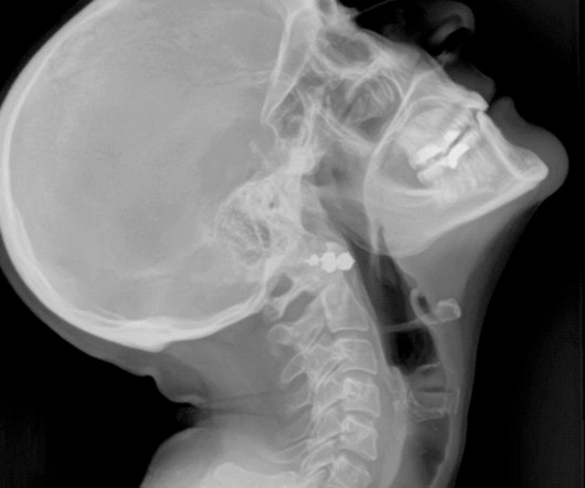
Closeup of X-ray photography of human brain Introduction: “The Radiant Thread” is a comprehensive guide that unravels the intricate world of X-ray imaging from A to Z. From its historical roots to contemporary innovations, we will follow the radiant thread that connects all aspects of X-ray imaging.
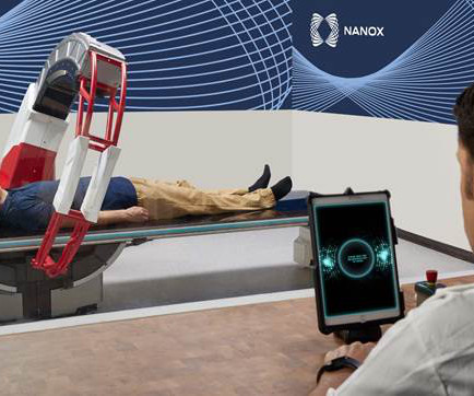
milla1cf Mon, 05/01/2023 - 17:39 May 1, 2023 — Nano-X Imaging Ltd. , Nanox.ARC is a stationary X-ray system intended to produce tomographic images of the human musculoskeletal system adjunctive to conventional radiography on adult patients.
Introduction: Radiology is a gateway to the invisible, offering a window into the human body and enabling a deeper understanding of health and disease. “X-Rays and Beyond” embarks on an exciting journey to explore the captivating world of radiology, highlighting how it goes far beyond just X-rays.
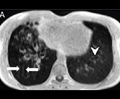
The authors of this study aimed to evaluate whether artificial intelligence, specifically a deep neural network (DNN), was able to distinguish between tuberculosis (TB) or nontuberculous mycobacterial lung disease (NTM-LD) patients through chest X-rays (CXRs) from suspected mycobacterial lung disease.
Helps Doctors Monitor Disease Progression Diagnostic imaging is one of the most important markers that doctors utilize to monitor the progression of diseases for a variety of ailments, including: Certain types of malignant cancer Pneumonia and other respiratory diseases Internal bleeding Brain injuries Cardiac conditions Acute injuries (i.e.,
In a retrospective study led by presenter Bin Zhang, MD, of The First Affiliated Hospital of Jinan University in China, 1,538 pairs of lumbar spine radiographs and dual-energy x-ray absorptiometry (DEXA) results from females aged ≥ 50 years were collected between January 1, 2014, and October 10, 2019.
Digital radiography utilizes a unique technology, similar to a digital camera, that instantly sends X-ray images to a computer for quick viewing. Likewise, digital radiography systems use a detector known as a flat panel to translate data into a digital electronic signal.
The new imaging systems that will be on display include three new digital radiography (DR) suites, two new fluoroscopy systems, a 0.4T Join Fujifilm in person at booth #1929 at RSNA and follow FUJIFILM Healthcare Americas Corporation on LinkedIn and X for #RSNA23 updates throughout the event. Will be available in the U.S.
Introduction: While X-rays are traditionally associated with skeletal imaging, their reach extends far beyond bones. Unveiling the Cardiovascular System: Angiography and Beyond: X-ray technology has revolutionized the visualization of the cardiovascular system.
S hamie Kumar describes her perspective on how radiography has evolved over time, the impact radiographers can have in detecting abnormal X-rays and reflects how she views fast approaching AI in advancing current skills. Do some radiographers add a text note on the X-ray itself?
Mobile imaging provides comprehensive X-Ray, EKG and ultrasound services directly to medical facilities,homesand businesses. When people hear the world mobile imaging, they typically think of portable X-Ray imaging. What is Mobile Imaging? Verify placement of other medical devices such as implantable pumps and catheters.
Closeup of X-ray photography of human brain Description: The field of radiology is in the midst of a remarkable revolution, driven by cutting-edge technologies and innovations. This aids in the early detection and monitoring of diseases.
Technology in Radiology : Radiology relies on a sophisticated array of imaging technologies, such as X-rays, CT scans, MRIs, and ultrasound, to reveal the complexities of human anatomy and pathology.
Absence of residual pneumothorax or the presence of an apical residual pneumothorax smaller than 2 cm, based on chest radiography. Chest X-rays were reviewed centrally by experts blind to the initial assessment, but these experts could likely tell if the device was a chest tube or aspiration device, which introduces bias.
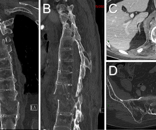
Ankylosing Spondylitis: Ankylosing spondylitis (AS) is a seronegative spondyloarthropathy and chronic inflammatory disease of the axial skeleton that leads to the partial or complete fusion and rigidity of the spine. 7 Primary diagnosis of AS involves radiography of sacroiliac joints and symptomatic areas of the spine (Figs.
Thompson is the radiography program director and associate professor for Austin Peay State University in Clarksville, Tennessee. My journey into the world of radiography started at 12 years old from a deeply personal place - accompanying my mother to medical appointments during her battle, which she sadly lost, with breast cancer.
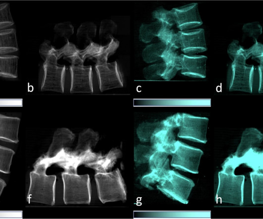
Dark-field x-ray imaging can provide insights into bone microstructure and could potentially have a role in assessing osteoporosis, according to a study published November 4 in European Radiology Experimental. Quantitative CT measurements can be used to screen for the disease. The donors' average age was 68.5 years
Likewise, it is in the best interest of the child to decline an order for an exam for scoliosis if you only have conventional radiography equipment and are incapable of offering the child the benefit of decreased exposure by standard PA positioning and digital imaging systems. Reference: 1 1 U.S.
X-rays , EKGs and ultrasounds are a foundational part of good healthcare because they help doctors diagnose diseases and develop treatment plans. It’s difficult, however, for many patients in a nursing home or other care environment to receive the appropriate diagnostic screenings in an efficient and comfortable manner.
According to the article, there is currently no ultrasound scanner or x-ray machine at the Obongi Health Centre IV, the main health facility in Obongi District in Uganda, and this is having a major impact on both the local population and the 130,000 refugees from South Sudan who are currently also served by the facility.
We organize all of the trending information in your field so you don't have to. Join 5,000 users and stay up to date on the latest articles your peers are reading.
You know about us, now we want to get to know you!
Let's personalize your content
Let's get even more personalized
We recognize your account from another site in our network, please click 'Send Email' below to continue with verifying your account and setting a password.


Let's personalize your content