This site uses cookies to improve your experience. To help us insure we adhere to various privacy regulations, please select your country/region of residence. If you do not select a country, we will assume you are from the United States. Select your Cookie Settings or view our Privacy Policy and Terms of Use.
Cookie Settings
Cookies and similar technologies are used on this website for proper function of the website, for tracking performance analytics and for marketing purposes. We and some of our third-party providers may use cookie data for various purposes. Please review the cookie settings below and choose your preference.
Used for the proper function of the website
Used for monitoring website traffic and interactions
Cookie Settings
Cookies and similar technologies are used on this website for proper function of the website, for tracking performance analytics and for marketing purposes. We and some of our third-party providers may use cookie data for various purposes. Please review the cookie settings below and choose your preference.
Strictly Necessary: Used for the proper function of the website
Performance/Analytics: Used for monitoring website traffic and interactions
66 year old female with abdominal pain. Diagnosis? • Xray of the Week Figure 1. What are the important findings? Figure 2. Axial CT - A: Large filling defect in the left atrium. Note the slight enhancement indicating it is a neoplastic mass rather than thrombus, (red arrow) suggesting myxoma. The large size is also compatible with myxoma rather than thrombus.
Diagnostic imaging is one of the best methods that medical practices, hospitals and health care professionals can utilize to diagnose and treat their patients. However, finding licensed and experienced radiologic technologists (rad techs) is becoming increasingly difficult. Why Trust AIMI’s Radiologic Technologists? Hiring has never been an easy process.
Breast screenings are integral to women’s’ health. They provide the critical information that healthcare providers rely on for breast cancer detection. Routine mammograms and other recommended breast screenings can help improve patient outcomes by paving the way for early detection. Here, we discuss the breast screening information all women should know.
Amidst rising cancer prevalence and soaring costs, new cancer technologies and innovations are emerging to support the early detection, treatment, and surveillance of cancer. Read this guide to understand how to evaluate these solutions for your employees and members – and to learn more about the current state of coverage, clinical and cost effectiveness, and impact on quality and outcomes.
A study published in the American Journal of Roentgenology highlights the role computed tomography (CT) has played in identifying electronic cigarette or vaping product use-associated lung injury, also known as EVALI. The first reports of EVALI emerged in late 2019. Within a six-month timeframe, a handful of cases reported to the Centers for Disease Control and Prevention (CDC) increased to nearly 3,000 and 68 confirmed deaths.
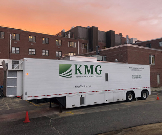
As hospitals and imaging facilities are figuring out the “new normal,” one thing remains the same: Making a capital equipment acquisition requires a well thought out plan. Many facilities are experiencing a surge in volume, larger than pre-pandemic numbers. And with the increase in patient demand many facilities are starting to focus on expansion and […] The post Financial Benefits with Mobile Imaging appeared first on Kings Medical Group.
As hospitals and imaging facilities are figuring out the “new normal,” one thing remains the same: Making a capital equipment acquisition requires a well thought out plan. Many facilities are experiencing a surge in volume, larger than pre-pandemic numbers. And with the increase in patient demand many facilities are starting to focus on expansion and […] The post Financial Benefits with Mobile Imaging appeared first on Kings Medical Group.
8 Year Old Male With Trauma Due To A Fall. Diagnosis? • Xray of the Week Figure 1. What is the important finding? Figure 2 A: AP view radiograph of right forearm. Acute transverse fracture of distal radius visualized (green arrow). B: Lateral radiograph view of right forearm. Angulation of ulna visualized (red arrows). Discussion: The above imaging findings occurred in an 8-year-old child with a trauma after a fall.
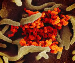
The structures of a virus can be elucidated by using the high resolving power of scanning electron microscopy. The post Electron Microscopy: What Does Coronavirus Look Like? appeared first on Open Medscience.
Breast augmentation is one of the most popular plastic surgery procedures performed today. This breast surgery is often performed long before a woman needs her first mammogram , which means that the topic of breast implants’ effects on necessary screenings is rarely discussed before breast augmentation is performed. If you’re nearing the age at which routine breast screenings become necessary (age 40), you may have some of the following common questions.
Patient-centric scheduling can only be achieved through optimized radiology workflows, effective communications between staff and physicians, and, of course, through specialized schedulers. In this guide, we’ll take you through a step-by-step process to transform your radiology center into a high-performance hub of medical imaging.
Dr Gareth Davies describes the massive impact the COVID 19 pandemic had on elective cross-sectional reporting, reducing output to almost zero. Here he reflects on how the drive for innovation and the motivation to think differently led to a better teleradiology service for both patients and staff. Dr Gareth Davies The pandemic will certainly define us as an organisation.
Mobile imaging is a huge—and growing—industry. Already worth more than $1.5 billion globally in 2016 , this is a market that is poised to grow rapidly in the years to come. We’d like to break down what it is about mobile imaging that makes it so valuable to healthcare organizations, and why you should take a close look at the pros of implementing mobile imaging as part of your operations.
42 year old male with headache and history of cerebral palsy. What is the diagnosis? • Xray of the Week Figure 1. Head CT. What are the significant findings? Figure 2. Head CT. Red arrows indicate focal encephalomalacia of the left parietal lobe with replacement by a cystic mass that communicates with the left lateral ventricle. Green arrows indicate white matter lining the porencephalic cyst.
PET imaging is used in oncology, neurology and cardiology. The post Molecular Imaging Technology: Use of PET in Clinical Microdose Studies appeared first on Open Medscience.
About 40% of us will be diagnosed with cancer in our lifetime, and patients are getting younger. At the same time, the cost of treatment continues to rise, with employers spending 8.5% more on cancer care for each employee than they did last year. The best thing employers can do for their employees and business tomorrow is to invest in cancer detection and care today.
90 yo F with UTI. Diagnosis? • Xray of the Week Figure 1. Non-contrast CT abdomen & pelvis of a 90-year-old female. Figure 2. Figure 1: Non-contrast CT abdomen & pelvis of a 90-year-old female with a UTI demonstrating emphysematous pyelonephritis and a renal stone within the renal pelvis. A. Coronal non-contrast CT of abdomen & pelvis showing gas (green arrow) and renal stone (red) within the renal pelvis.
Name the cardiac device. • Xray of the Week Figure 1. Figure 1. AP Chest X-ray demonstrating placement of 3 MitraClips over the tricuspid valve. Figure 2. A: AP chest X-ray demonstrating placement of 3 MitraClips over the tricuspid valve (yellow arrow) B: Coronal chest CT showing 3 MitraClips over the tricuspid valve in the right atrioventricular septum (red arrow).
A 67 year old female with trauma due to a motor vehicle collision. What is the diagnosis? • Xray of the Week Figure 1. Brain CT. What is the significant finding. Figure 2. Axial CT brain demonstrates a right posterior lens dislocation with the lens lying in the dependent portion of the vitreous humor (red arrow). Note the normal location of the left lens in the iris (green arrow).
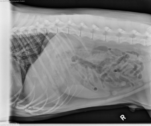
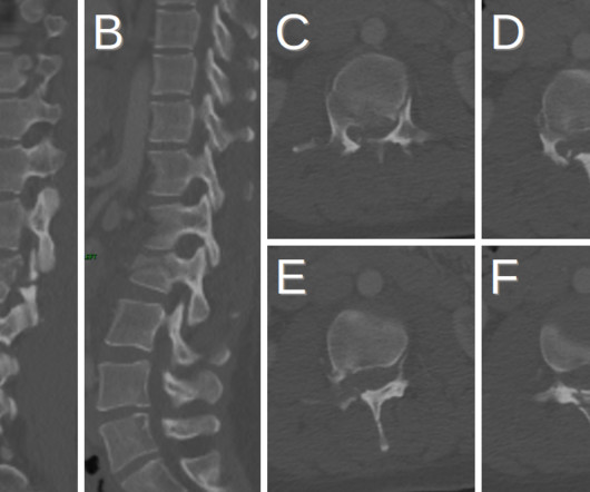
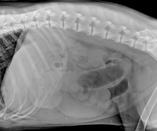
Trauma in a 31 yo F due to a motor vehicle collision (MVC) • Xray of the Week Figure 1. What are the important findings on this CT scan. What is the diagnosis? Figure 2. A,B. Sagittal reformatted CT images. C-E. Axial CT images Images show horizontal fracture through the right lamina (orange arrows), right pedicle (green arrows) and left pedicle (red arrows).
Discover how Color's comprehensive care solution is revolutionizing cancer screening adherence and knowledge. Through an in-depth case study, Color's unique approach to comprehensive cancer care has shown significant benefits in increasing screening rates and enhancing patient knowledge. Participants reported a 2-3x increase in adherence to screening guidelines over just 8 weeks, with 84% of participants increasing their familiarity with timing and frequency of cancer screening.
We organize all of the trending information in your field so you don't have to. Join 5,000 users and stay up to date on the latest articles your peers are reading.
You know about us, now we want to get to know you!
Let's personalize your content
Let's get even more personalized
We recognize your account from another site in our network, please click 'Send Email' below to continue with verifying your account and setting a password.


Let's personalize your content