Artificial Intelligence Technology Advances Heart Imaging
Health IT Analytics
SEPTEMBER 2, 2021
New artificial intelligence… read more

Health IT Analytics
SEPTEMBER 2, 2021
New artificial intelligence… read more
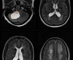
Dr Balaji Anvekar FRCR
AUGUST 29, 2021
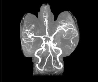
Clinical Details: middle-aged female, altered sensorium after convulsions. MRI study of brain shows: Abnormal T2 hyperintensity with marked focal parenchymal swelling due to vasogenic oedema involving left temporal, insular cortex and adjacent opercular parietal white matter. Diffusion restriction in corresponding region confined to cortical grey matter of left temporal lobe and adjacent insular cortex on diffusion weighted images.
This site is protected by reCAPTCHA and the Google Privacy Policy and Terms of Service apply.
Lake Medical Imaging
SEPTEMBER 2, 2021
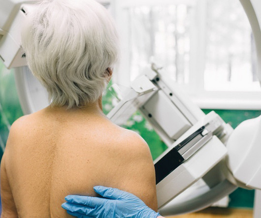
Senior woman having a mammography scan Breast density is associated with an increased risk of invasive cancer among women aged 65 and up, according to a new prospective study published in JAMA Network Open.Meanwhile, less-dense, fatty tissue was tied to a lower probability of this form of the disease across all age groups. Research [.

Premier Radiology Services
SEPTEMBER 1, 2021
The Need For an Error-Proof Workspace The biggest expense most radiologists have is malpractice insurance, and it is very important to be able to work in an error-proof environment in order to give the best patient care. Reading Radiologists carry a lot of responsibility on their shoulders. You need to be focused, organized, and provide great care and attention to every scan that comes across your screen.
Advertisement
Amidst rising cancer prevalence and soaring costs, new cancer technologies and innovations are emerging to support the early detection, treatment, and surveillance of cancer. Read this guide to understand how to evaluate these solutions for your employees and members – and to learn more about the current state of coverage, clinical and cost effectiveness, and impact on quality and outcomes.


Health IT Analytics
SEPTEMBER 1, 2021
To improve population… read more
Diagnostic Imaging Brief brings together the best content for architecture professionals from the widest variety of industry thought leaders.

Global Radiology CME
SEPTEMBER 1, 2021
We are pleased to announce Global Radiology CME's Kevin Rice, MD is a semifinalist for 2021 ,AuntMinnie.com 's Most Effective Radiology Educator. Dr. Kevin Rice, Vice Chief of the Medical Staff at Valley Presbyterian Hospital, a 21 year radiologist at RIMA, a proud member of Radiology Partners, a member of the Radiology Partners Advocacy Board, and President of Global Radiology CME, is honored to be named a semi finalist in the " 2021 edition of the Minnies, AuntMinnie.com's campaign to recogniz

Pure Mammography
AUGUST 30, 2021
Lumps in the breasts are typically believed to be a cause for great concern, although studies indicate that approximately 60 percent of women have them. In some cases, lumps are concerning, but not always. Here, we discuss the term “fibrocystic breast changes” and what it may mean for women with lumpy or ropy breast tissue. FIbrocystic breasts are described as having a particular texture.

Health IT Analytics
SEPTEMBER 1, 2021
In a cohort study,… read more
Neuroradiology Cases
AUGUST 29, 2021
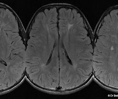
Ears of the lynx sign The ears of the lynx sign refers to an abnormal bilateral symmetric cone-shaped hyperintensity on FLAIR and T2w images at the tip of the frontal horns of lateral ventricles. The abnormality corresponds to the region of forceps minor which resembles the tufts of hair crowning the ears of a lynx. Sagittal T1w images show an associated thin stripe of corpus callosum.

Advertisement
Patient-centric scheduling can only be achieved through optimized radiology workflows, effective communications between staff and physicians, and, of course, through specialized schedulers. In this guide, we’ll take you through a step-by-step process to transform your radiology center into a high-performance hub of medical imaging.


Health IT Analytics
AUGUST 30, 2021
Data analytics serves as an… read more

Health IT Analytics
AUGUST 31, 2021
University of Los Angeles… read more

Health IT Analytics
AUGUST 30, 2021
According to a new study,… read more

Expert insights. Personalized for you.


Let's personalize your content