This site uses cookies to improve your experience. To help us insure we adhere to various privacy regulations, please select your country/region of residence. If you do not select a country, we will assume you are from the United States. Select your Cookie Settings or view our Privacy Policy and Terms of Use.
Cookie Settings
Cookies and similar technologies are used on this website for proper function of the website, for tracking performance analytics and for marketing purposes. We and some of our third-party providers may use cookie data for various purposes. Please review the cookie settings below and choose your preference.
Used for the proper function of the website
Used for monitoring website traffic and interactions
Cookie Settings
Cookies and similar technologies are used on this website for proper function of the website, for tracking performance analytics and for marketing purposes. We and some of our third-party providers may use cookie data for various purposes. Please review the cookie settings below and choose your preference.
Strictly Necessary: Used for the proper function of the website
Performance/Analytics: Used for monitoring website traffic and interactions
A study published in the American Journal of Roentgenology highlights the role computed tomography (CT) has played in identifying electronic cigarette or vaping product use-associated lung injury, also known as EVALI. The first reports of EVALI emerged in late 2019. Within a six-month timeframe, a handful of cases reported to the Centers for Disease Control and Prevention (CDC) increased to nearly 3,000 and 68 confirmed deaths.
A 67 year old female with trauma due to a motor vehicle collision. What is the diagnosis? • Xray of the Week Figure 1. Brain CT. What is the significant finding. Figure 2. Axial CT brain demonstrates a right posterior lens dislocation with the lens lying in the dependent portion of the vitreous humor (red arrow). Note the normal location of the left lens in the iris (green arrow).
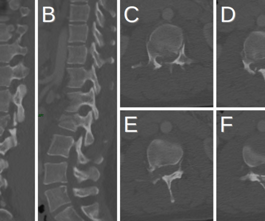
Trauma in a 31 yo F due to a motor vehicle collision (MVC) • Xray of the Week Figure 1. What are the important findings on this CT scan. What is the diagnosis? Figure 2. A,B. Sagittal reformatted CT images. C-E. Axial CT images Images show horizontal fracture through the right lamina (orange arrows), right pedicle (green arrows) and left pedicle (red arrows).
We organize all of the trending information in your field so you don't have to. Join 5,000 users and stay up to date on the latest articles your peers are reading.
You know about us, now we want to get to know you!
Let's personalize your content
Let's get even more personalized
We recognize your account from another site in our network, please click 'Send Email' below to continue with verifying your account and setting a password.


Let's personalize your content