This site uses cookies to improve your experience. To help us insure we adhere to various privacy regulations, please select your country/region of residence. If you do not select a country, we will assume you are from the United States. Select your Cookie Settings or view our Privacy Policy and Terms of Use.
Cookie Settings
Cookies and similar technologies are used on this website for proper function of the website, for tracking performance analytics and for marketing purposes. We and some of our third-party providers may use cookie data for various purposes. Please review the cookie settings below and choose your preference.
Used for the proper function of the website
Used for monitoring website traffic and interactions
Cookie Settings
Cookies and similar technologies are used on this website for proper function of the website, for tracking performance analytics and for marketing purposes. We and some of our third-party providers may use cookie data for various purposes. Please review the cookie settings below and choose your preference.
Strictly Necessary: Used for the proper function of the website
Performance/Analytics: Used for monitoring website traffic and interactions
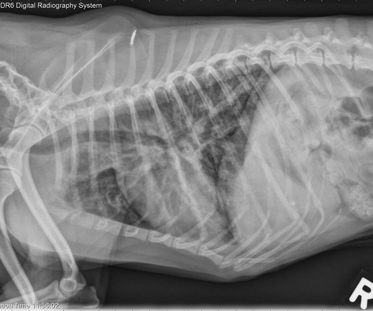
59-year-old male with trauma. Diagnosis? • Xray of the Week Figure 1. What are the important findings? Figure 2. A: Sagittal CT image demonstrates the right posterior lens subluxation with the inferior portion of the lens displaced posteriorly into the vitreous humor (red arrow). B: Sagittal CT image demonstrates the normal location of the left lens in the iris (green arrow).
Ireland welcomed back vaccinated tourists just under 2 weeks ago and Dublin is returning to normal. Take a live peek at The Temple Bar Pub in Dublin, just a 5 minute walk from the Global Radiology conference venue, The Westin Dublin Hotel. As of July 31, 2021 over 86% of adult population in Ireland have received at least one dose and 71% of eligible adults are now fully vaccinated.
66 year old female with abdominal pain. Diagnosis? • Xray of the Week Figure 1. What are the important findings? Figure 2. Axial CT - A: Large filling defect in the left atrium. Note the slight enhancement indicating it is a neoplastic mass rather than thrombus, (red arrow) suggesting myxoma. The large size is also compatible with myxoma rather than thrombus.
Amidst rising cancer prevalence and soaring costs, new cancer technologies and innovations are emerging to support the early detection, treatment, and surveillance of cancer. Read this guide to understand how to evaluate these solutions for your employees and members – and to learn more about the current state of coverage, clinical and cost effectiveness, and impact on quality and outcomes.
We organize all of the trending information in your field so you don't have to. Join 5,000 users and stay up to date on the latest articles your peers are reading.
You know about us, now we want to get to know you!
Let's personalize your content
Let's get even more personalized
We recognize your account from another site in our network, please click 'Send Email' below to continue with verifying your account and setting a password.


Let's personalize your content