Artificial Intelligence Advances Breast Cancer Detection
Health IT Analytics
OCTOBER 7, 2021
With artificial… read more

Health IT Analytics
OCTOBER 7, 2021
With artificial… read more

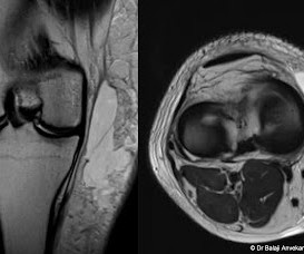
Dr Balaji Anvekar FRCR
OCTOBER 3, 2021
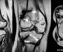
Clinically young male patient with athletic background complaining of typical unilateral anterior knee pain. Marked tenderness at the superior pole of patella. This MRI study of knee joint shows abnormal irregularity, fragmentation with sclerosis involving superior pole of patella. An associated thickening of quadriceps tendon. Knee joint effusion. Imaging findings consistent with osteochondrosis of patella at superior pole.
This site is protected by reCAPTCHA and the Google Privacy Policy and Terms of Service apply.
Neuroradiology Cases
OCTOBER 8, 2021
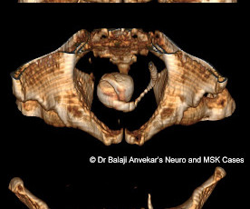
This non-contrast CT study of abdomen for KUB of patient with left abdominal pain shows a dense large calculus in Urinary bladder which is in continuity with ribbon like column of multiple calculi in left ureter. Retrograde history taking unfurled that the patient had undergone ESWL one month ago. Imaging diagnosis: Steinstrasse. Steinstrasse is the German term which means "stone street", used to describe a possible complication of extracorporeal shock wave lithotripsy (ESWL) for urinary tract c

Premier Radiology Services
OCTOBER 7, 2021
Teleradiology Reading Platform Agreement The teleradiology services provider delivers a high availability solution and customized workflows through its partnership with RamSoft. The announcement of Teleradiology Reading Platform agreement was made ahead of the Radiological Society of North America (RSNA) Annual Meeting in Chicago. Learn more about the Premier Radiology Services case study at RSNA Booth #4106.

Advertisement
Amidst rising cancer prevalence and soaring costs, new cancer technologies and innovations are emerging to support the early detection, treatment, and surveillance of cancer. Read this guide to understand how to evaluate these solutions for your employees and members – and to learn more about the current state of coverage, clinical and cost effectiveness, and impact on quality and outcomes.


Health IT Analytics
OCTOBER 5, 2021
According to a study by the… read more
Diagnostic Imaging Brief brings together the best content for architecture professionals from the widest variety of industry thought leaders.

Associates in Medical Imaging
OCTOBER 6, 2021
For healthcare organizations, achieving positive patient outcomes is the top priority. Medical imaging plays a key role in making a diagnosis and determining the right course of treatment. Large hospital systems are often looking for ways to offer diagnostic capabilities to all satellite locations, bringing the convenience of imaging right to their patients.
Neuroradiology Cases
OCTOBER 3, 2021
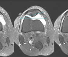
This MRI Axial STIR sections of knee show clinical marker on skin on anteromedial aspect of knee joint. There is patellar tilt, articulating surface of patella facing medially, abnormal thickening of lateral patellar retinaculum and patellofemoral ligament. Associated bone marrow oedema involving lateral margin of lateral articulating facet of patella.

Health IT Analytics
OCTOBER 7, 2021
According to the results of… read more
Dr Balaji Anvekar FRCR
OCTOBER 2, 2021
Clinical details, discharge summary mentions corrective osteotomy done for congenital progressive uni lateral knee deformity. Previous imaging details not available. This MRI of knee joint shows post-operative status with corrective osteotomy for femur and tibia. Well defined bone signal intensity outgrowth with cortex and medulla involving medial epiphysis of distal end of femur, epiphysis of tibial tuberosity with distinct cortex, medulla and fatty marrow in continuity with parent bone.

Advertisement
Patient-centric scheduling can only be achieved through optimized radiology workflows, effective communications between staff and physicians, and, of course, through specialized schedulers. In this guide, we’ll take you through a step-by-step process to transform your radiology center into a high-performance hub of medical imaging.

Dr Balaji Anvekar FRCR
OCTOBER 2, 2021
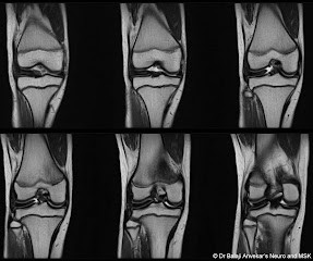
This MRI study of knee joint depicts discoid lateral meniscus. No obvious associated meniscal tear or para meniscal cyst. Discoid meniscus This is a congenital condition and is bilateral in about 50% of the cases. Usually encountered as an incidental finding on MRI examination in about 5% of the cases, typically affecting lateral meniscus. Discoid medial meniscus is very rare.

Health IT Analytics
OCTOBER 4, 2021
According to UT… read more

Health IT Analytics
OCTOBER 6, 2021
Regenstrief Institute… read more

Health IT Analytics
OCTOBER 4, 2021
Those managing chronic… read more

Advertisement
About 40% of us will be diagnosed with cancer in our lifetime, and patients are getting younger. At the same time, the cost of treatment continues to rise, with employers spending 8.5% more on cancer care for each employee than they did last year. The best thing employers can do for their employees and business tomorrow is to invest in cancer detection and care today.
Neuroradiology Cases
OCTOBER 3, 2021
Clinically RTA, run over by tractor. This MRI study shows a focal well defined lentiform shaped subcutaneous collection on medial aspect of knee joint superficial to the superficial fascia. Collection is clear, hypo intense on T1-weighted images without any septations or loculations. No obvious high signal intensity methaemoglobin staining on T1-weighted images to suggest any frank haematoma.

Neuroradiology Cases
OCTOBER 2, 2021
Clinically young patient presented with anteromedial knee pain. MRI sagittal T2 and axial T2 images delineates a linear well-defined low signal intensity band running across medial patellofemoral recess. However, there is no obvious associated bone marrow oedema involving medial articulating facet of patella or medial femoral trochlea. Mild associated joint effusion.

Neuroradiology Cases
OCTOBER 2, 2021
Clinically severe painful restricted shoulder movement, especially the abduction. Patient complaining that symptoms got aggravated over the period of time with shoulder exercise and physiotherapy. No obvious history of arthroscopy or intra articular injection. This MRI shoulder joint shows a well-defined tear drop shaped cystic lesion along supraspinatus tendon tapering laterally towards its insertion suggestive of intra tendinous ganglion cyst.

Expert insights. Personalized for you.


Let's personalize your content