This site uses cookies to improve your experience. To help us insure we adhere to various privacy regulations, please select your country/region of residence. If you do not select a country, we will assume you are from the United States. Select your Cookie Settings or view our Privacy Policy and Terms of Use.
Cookie Settings
Cookies and similar technologies are used on this website for proper function of the website, for tracking performance analytics and for marketing purposes. We and some of our third-party providers may use cookie data for various purposes. Please review the cookie settings below and choose your preference.
Used for the proper function of the website
Used for monitoring website traffic and interactions
Cookie Settings
Cookies and similar technologies are used on this website for proper function of the website, for tracking performance analytics and for marketing purposes. We and some of our third-party providers may use cookie data for various purposes. Please review the cookie settings below and choose your preference.
Strictly Necessary: Used for the proper function of the website
Performance/Analytics: Used for monitoring website traffic and interactions
Every field of medicine strives to innovate, improve, and demand high standards of excellence from its practitioners. This is why American physicians and health care providers stand out in the world of medicine. The field of radiology is no different. We serve patients and physicians to improve diagnostics, treatments, and recovery. We are at the center of patient care and we take that responsibility very seriously.
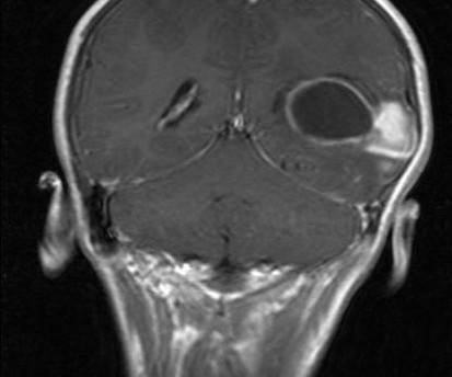
History – A 14 yr female c/o headache since 2 months. 2 episodes of seizures in last one week Findings- An ill-defined soild-cystic mass is seen in the left parietal lobe with surrounding mild to moderate edema. The cystic component shows restriction of diffusion and rim enhancement. The solid component shows avid postcontrast enhancement Final Diagnosis- Pleomorphic xanthoastrocytoma (PXA) Golden Point DDs of cyst with mural nodule- Pilocytic astrocytoma, PXA, ganglioglioma, hemangioblastoma, c
For the third year in a row, a Capitol Imaging Services affiliate has been designate as the best of the best! Our Southeast Louisiana network member, Diagnostic Imaging Services (DIS), was chosen by the readers of EDGE Magazine published in St. Tammany Parish. Diagnostic Imaging Services has been serving the greater New Orleans metropolitan market, the state of Louisiana and the Gulf South for nearly 50 years.
Africa is the world’s second largest and second-most populous continent , after Asia in both cases. At about 30.3 million km2 (11.7 million square miles) including adjacent islands, it covers 6% of Earth’s total surface area and 20% of its land area. With 1.38 billion people as of November 2021, it accounts for about 16.72% of the world’s human population.
Amidst rising cancer prevalence and soaring costs, new cancer technologies and innovations are emerging to support the early detection, treatment, and surveillance of cancer. Read this guide to understand how to evaluate these solutions for your employees and members – and to learn more about the current state of coverage, clinical and cost effectiveness, and impact on quality and outcomes.
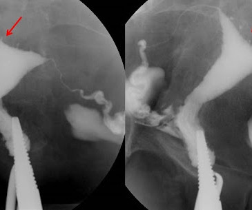
History: A 27 years female c/o menorrhagia. HSG is shown below. Findings: HSG study shows multiple small outpouchings from the uterine cavity, especially in the fundus The uterine cavity is otherwise preserved The tubes are normal with free spill Findings are s/o adenomyosis Extra Edge Point : Investigation of choice and findings--MRI – Widening of the junctional zone >12 mm, multiple small cystic lesions, pseudodiverticulae Famous Radiology Blog by Dr Sumer Sethi www.sumersethi.
History: A 27 years female c/o menorrhagia. HSG is shown below. Findings: HSG study shows multiple small outpouchings from the uterine cavity, especially in the fundus The uterine cavity is otherwise preserved The tubes are normal with free spill Findings are s/o adenomyosis Extra Edge Point : Investigation of choice and findings--MRI – Widening of the junctional zone >12 mm, multiple small cystic lesions, pseudodiverticulae Famous Radiology Blog by Dr Sumer Sethi www.sumersethi.
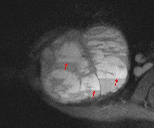
History A 13 years male c/o pain and swelling in right shoulder Findings: The Xrays shows a large expansile lytic lesion is seen in the right distal clavicle with a large associated soft tissue component in an immature skeleton The MRI shows a large expansile lytic lesion is seen in the right distal clavicle Multiple fluid-fluid levels are seen throughout the lesion Findings are typical of ABC of clavicle Diagnosis Aneurysmal Bone Cyst Famous Radiology Blog by Dr Sumer Sethi www.sumersethi.
Xray- Frontal radiograph of wrist shows multiple osteolytic areas with surrounding sclerosis is seen involving all carpal bones and second metatcarpal bone s/o permeative type of bone destruction. MRI- MRI shows Extensive areas of altered marrow signal with osteolysis are seen involving all the carpal bones and almost the entire 2nd metacarpal bone.
There are many confusing terms surrounding healthcare, healthcare services and health insurance coverage. Numerous different options can often put someone’s head in a spin as they try to grasp with bills and statements from medical provider offices, hospitals and medical centers. One term that Capitol Imaging Services often uses and can be considered to be a major financial benefit to people undergoing diagnostic scans is called “global billing.” In its simplistic form, global
We organize all of the trending information in your field so you don't have to. Join 5,000 users and stay up to date on the latest articles your peers are reading.
You know about us, now we want to get to know you!
Let's personalize your content
Let's get even more personalized
We recognize your account from another site in our network, please click 'Send Email' below to continue with verifying your account and setting a password.


Let's personalize your content