This site uses cookies to improve your experience. To help us insure we adhere to various privacy regulations, please select your country/region of residence. If you do not select a country, we will assume you are from the United States. Select your Cookie Settings or view our Privacy Policy and Terms of Use.
Cookie Settings
Cookies and similar technologies are used on this website for proper function of the website, for tracking performance analytics and for marketing purposes. We and some of our third-party providers may use cookie data for various purposes. Please review the cookie settings below and choose your preference.
Used for the proper function of the website
Used for monitoring website traffic and interactions
Cookie Settings
Cookies and similar technologies are used on this website for proper function of the website, for tracking performance analytics and for marketing purposes. We and some of our third-party providers may use cookie data for various purposes. Please review the cookie settings below and choose your preference.
Strictly Necessary: Used for the proper function of the website
Performance/Analytics: Used for monitoring website traffic and interactions
Women’s imaging at this year’s RSNA reflects momentous changes in trends regarding personalized, targeted healthcare in 2024 for women’s health. Research efforts toward supplemental breast imaging have ramped up in recent years and in Chicago, attendees can see results from these imagingmodalities being put to the test.
Ultrasound's utility will be on full display at this year's RSNA annual meeting, showing its merit in a wide variety of clinical applications. Research to be presented at the annual meeting in Chicago will explore the modality's clinical applications in musculoskeletal, pediatric, abdominal, and women's imaging among other areas.
An immersive virtual reality (VR)-based test can distinguish between operators with varying levels of thoracic ultrasound skill, a Danish study published January 6 in Ultrasound in Medicine & Biology found. With ultrasound being a user-dependent imagingmodality, operator skills are important for diagnostic accuracy.
Studies to be presented in Chicago will explore ultrasound’s clinical applications in musculoskeletal, pediatric, abdominal, and women's imaging among other applications. Furthermore, ultrasound's use as a supplemental tool will be explored, including for breast cancer detection and follow-up imaging. 11:00 a.m. |
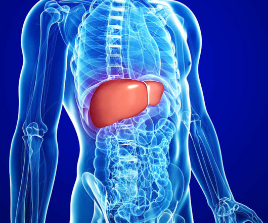
Clinicians need education to understand hepatocellular carcinoma (HCC) tracking and surveillance resources, according to research published May 14 in JAMA Network Open. This includes a perceived lack of diagnostic radiology resources, with ultrasound being the go-to imagingmodality for liver surveillance.
ChatGPT demonstrates modest accuracy when assigning BI-RADS scores for mammograms and breast ultrasound exams, according to research published October 30 in Clinical Imaging. Previous reports suggest that large language models can correctly recommend appropriate imagingmodalities for patients based on their clinical presentation.
A large variety of methods and modalities will be showcased for breast cancer screening, diagnosis, and treatment, as well as other pathologies. Not surprisingly, there will also be no shortage of presentations on the use of AI for breast imaging applications. However, there’s more to women’s imaging than breast imaging.
"Radiologists are uniquely positioned to educate other healthcare providers on how to properly remove personal health information from radiologic imaging files," wrote Stern and colleague William Weadock, MD, also of the university. The investigators conducted a study that followed previous work Weadock published 15 years ago.
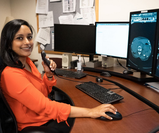
Becoming a radiologist requires an extraordinary level of dedication, education, and training. Radiologists are medical doctors who specialize in interpreting imaging studies like X-rays, CT scans, MRIs, and ultrasounds to diagnose and guide treatment for various conditions.
To excel in medical imaging, one must pursue postgraduate education beyond basic nursing or medicine. The post A Guide To Education and Upskilling For Professionals in The Medical Imaging Field appeared first on Open Medscience.
There are plenty of resources available for education and support around pediatric imaging. Here are a few that are particularly useful no matter where you find yourself imaging children. You can enroll in the free, on-demand course at Pediatric Appendix Ultrasound Standardized Performance and Reporting Training.
Section 1: The Shifting Paradigm in Trauma Imaging: Introduce the changing dynamics in trauma radiology, highlighting the transition from traditional imaging approaches to the emergence of extraneous imagingmodalities.
Discuss iterative reconstruction techniques that allow for lower radiation doses without compromising image quality. Low-Dose ImagingModalities: A Shift Towards Safety: Introduce low-dose imagingmodalities as a safer alternative.
This blog post explores recent innovations in cardiac imaging technologies, shedding light on advancements that are shaping the landscape of cardiovascular healthcare. Functional MRI (fMRI) for Dynamic Insights: Mapping Real-Time Changes in Cardiac Activity: Highlight the role of functional MRI in cardiac imaging.
Advances in Ultrasound Technology: Non-invasive and Radiation-Free Imaging: Explore recent advances in pediatric ultrasound technology. Discuss how ultrasound, being non-invasive and radiation-free, is increasingly used in pediatric imaging for various conditions.
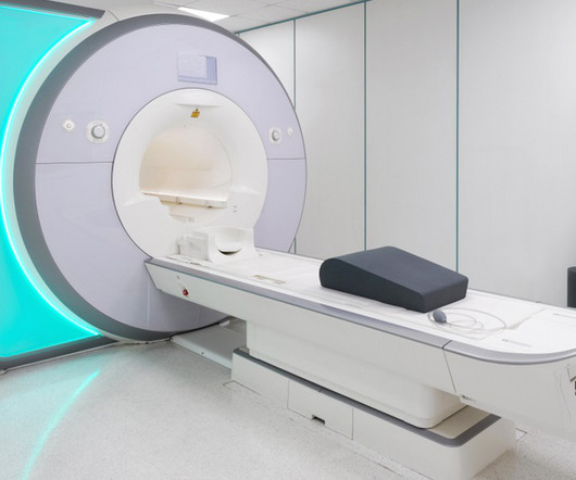
Technology in Radiology : Radiology relies on a sophisticated array of imaging technologies, such as X-rays, CT scans, MRIs, and ultrasound, to reveal the complexities of human anatomy and pathology.
Introduction to Radiology : Radiology is a branch of medicine that uses medical imaging techniques to diagnose and treat diseases and injuries. It includes various imagingmodalities such as X-rays, CT scans, MRIs, ultrasounds, and nuclear medicine.
Radiography has exploded into a variety of modalities and specialisms from CT to Ultrasound to MRI; all driven by research and development. I’m also passionate about education, research, and (of course) radiography. That is where I come in. That is to say, I’m fairly easy to pick out of a line up.
Patient communication and education may be key for radiologists in the wake of an advisory from the U.S. Radiology already has well-defined roles in imaging some of these cancers, specifically breast, liver, and head and neck cancer. Surgeon General, Vivek Murthy, MD,on alcohol-related cancer risk.
The theoretical basis for ultrasound physics has been around since 1794, but it wasn’t until 1942, when Dr Karl Theodore Dussik in Austria transmitted an ultrasound beam through a human skull to view the brain, that ultrasound was first used in medicine. (13) This was a defining publication in the field of medical ultrasound. (14)
Magnetic resonance–guided focused ultrasound thalamotomy restored distinctive resting-state networks in patients with essential tremor. JNS22411 Magnetic resonance–guided focused ultrasound (MRgFUS) thalamotomy ameliorates symptoms in patients with essential tremor (ET). Kato S, Maesawa S, Bagarinao E, et al. J Neurosurg.
Since then, the modality has helped reveal the brain's structural and functional activity and the effects of life-threatening events such as stroke. It has also improved the diagnosis of cancer, played a leading role in identifying musculoskeletal injuries, and added value as an adjunctive breast imagingmodality. 12:15 p.m. |
We organize all of the trending information in your field so you don't have to. Join 5,000 users and stay up to date on the latest articles your peers are reading.
You know about us, now we want to get to know you!
Let's personalize your content
Let's get even more personalized
We recognize your account from another site in our network, please click 'Send Email' below to continue with verifying your account and setting a password.


Let's personalize your content