Remove Emergency Room Remove Events Remove Imaging
article thumbnail

Interstitial lung abnormalities linked to respiratory disease risk

AuntMinnie

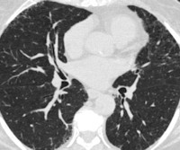
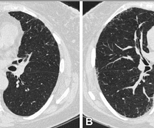
CT imaging shows that severe acute respiratory disease events can be caused by quantitative interstitial abnormalities (QIA) -- that is, small irregularities that don't necessarily meet diagnostic criteria for advanced pulmonary diseases but show up on CT exams over time, a study published April 30 in Radiology has reported.

Disease 111
article thumbnail

Lung Abnormality Progression Linked to Acute Respiratory Disease in Smokers

Imaging Technology

milla1cf Fri, 05/24/2024 - 07:00 May 24, 2024 — Smokers who have small abnormalities on their CT scans that grow over time have a greater likelihood of experiencing acute respiratory disease events, according to a new study published today in Radiology , a journal of the Radiological Society of North America ( RSNA ).

Disease 94
Insiders

Sign Up for our Newsletter

This site is protected by reCAPTCHA and the Google Privacy Policy and Terms of Service apply.

Trending Sources

article thumbnail

CHRISTUS Health Hits Texas Roads

Imaging Technology

Luckily, when they happen in East Texas, fast access to medical care is possible at many sporting events thanks to CHRISTUS Health ’s Mobile Athletic Training Room (MATR) — parked just steps away from the playing field. FDR AQRO provides instant imaging and answers just steps away from the field.

X-ray 126
article thumbnail

Overnight Insights: The Advantage of Night Hawk Radiology in Critical Cases (Tamil Nadu, Sivaganga, Thanjavur, Theni, Thoothukudi)

Future Teleradiology

Immediate Diagnostic Insights: Discuss the pivotal role of Night Hawk Radiology in providing immediate image interpretations, particularly in emergency and critical care scenarios, and the advantage it brings to such cases. Section 1: Introduction to Night Hawk Radiology 1.1 Section 2: The Advantage in Critical Cases 2.1

article thumbnail

Can MRI Diagnose Dementia?

The Radiology Clinic

An MRI is a high-resolution imaging tool that can reveal structural abnormalities within the body. They appear dark on this specific image type. On the left image above, you can see an example of what normal ventricles look like. In the image below you can see white spots throughout the brain.

article thumbnail

Managing the Patient with Alcohol Intoxication

REBEL EM

Klein 2018) Despite the high risk of pathology and decompensation, many patients with presumed alcohol intoxication can be evaluated and discharged without any labs or imaging. Any report of trauma should prompt evaluation with imaging. Any facial or scalp lacerations, abrasions, ecchymosis, or deformities should prompt imaging.

article thumbnail

AuntMinnie 2001: Frontline radiologists recall September 11

AuntMinnie

Perhaps the most tragic aspect of their stories is that the PACS networks, disaster protocols, and emergency rooms were not severely strained, since most of the victims did not survive. In the following accounts, radiology professionals who were on the front lines discuss the events of September 11, 2001.