This site uses cookies to improve your experience. To help us insure we adhere to various privacy regulations, please select your country/region of residence. If you do not select a country, we will assume you are from the United States. Select your Cookie Settings or view our Privacy Policy and Terms of Use.
Cookie Settings
Cookies and similar technologies are used on this website for proper function of the website, for tracking performance analytics and for marketing purposes. We and some of our third-party providers may use cookie data for various purposes. Please review the cookie settings below and choose your preference.
Used for the proper function of the website
Used for monitoring website traffic and interactions
Cookie Settings
Cookies and similar technologies are used on this website for proper function of the website, for tracking performance analytics and for marketing purposes. We and some of our third-party providers may use cookie data for various purposes. Please review the cookie settings below and choose your preference.
Strictly Necessary: Used for the proper function of the website
Performance/Analytics: Used for monitoring website traffic and interactions
“So we were thinking and asking ourselves, ‘can nonradiologists benefit from AI and chest radiography analysis in this emergency unit set.’ ” Per year, LMU receives between 5,000 and 6,000 orders for chest radiographs for primary diagnosis from the emergency unit alone.
The green project conducted by radiographers at the European Institute of Oncology in Milan and nearby Hospital of Legnano saved an estimated 12,000 euros ($13,000), said Andrea Masperi, who presented the details. The group developed a "green imaging review" for patients accessing the emergencyroom.
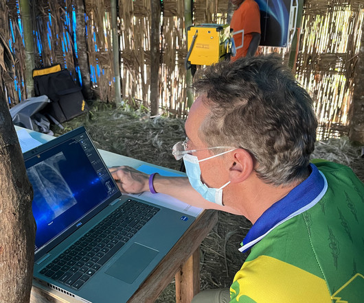
Rob Liddell, MD, is a diagnostic radiologist who used MinXray’s Impact Wireless system to take radiographic images of patients in the village and screen for common diseases in the region, such as tuberculosis and emphysema. He also used the system to diagnose cancers, infections and various musculoskeletal injuries.
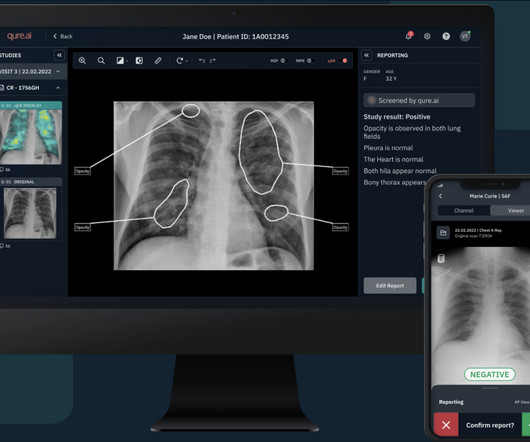
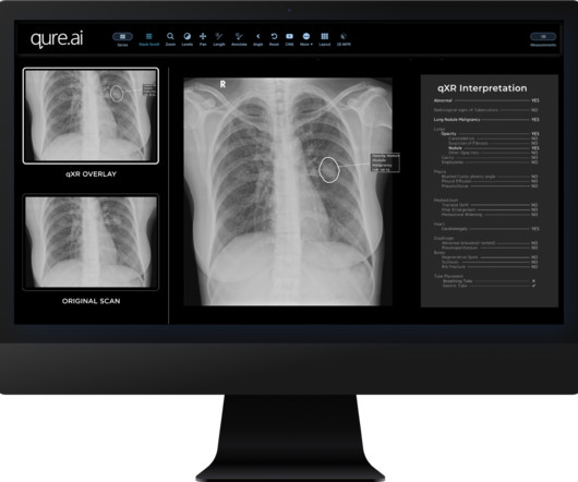
It can also serve as a crucial second reader for physicians, assisting in the review of frontal (AP/PA) chest radiographs of adults acquired on digital radiographic systems. The performance of various readers, including radiologists, pulmonologists, and emergencyroom physicians, showed improvement.
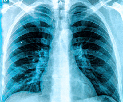
We are excited about the ability to deploy this in emergencyroom, inpatient and intensive care medicine. “Using our AI-based software and DDR, we can generate digital PFT's (dPFTs) which might be used as surrogates for pulmonary function testing. Chest radiography is typically acquired in the evaluation of pulmonary disorders.
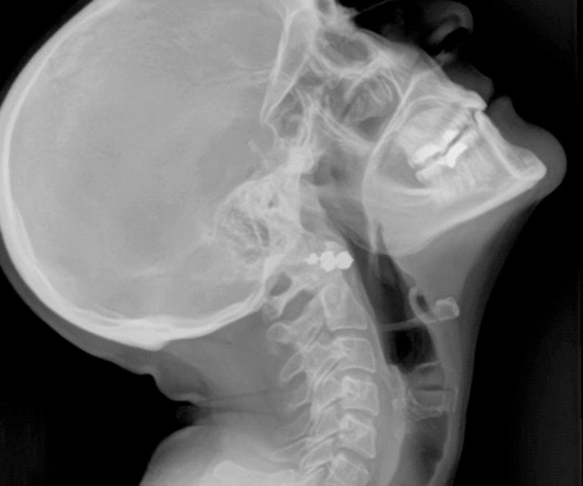
Yes, there is an acute shortage of manpower in this space and if recent reports are anything to go by, the volume of radiographic procedures being conducted will surpass the number of radiologist being inducted into the field by a ratio of 15:2. A patient in his mid-40s had the worst headache of his life. It was around 11.30
Qure’s qXR has now been cleared to triage pneumothorax (PTX) and pleural effusion (PE), which present severe challenges in emergencyrooms and intensive care units (ICUs), according to the company statement released today.
We organize all of the trending information in your field so you don't have to. Join 5,000 users and stay up to date on the latest articles your peers are reading.
You know about us, now we want to get to know you!
Let's personalize your content
Let's get even more personalized
We recognize your account from another site in our network, please click 'Send Email' below to continue with verifying your account and setting a password.


Let's personalize your content