This site uses cookies to improve your experience. To help us insure we adhere to various privacy regulations, please select your country/region of residence. If you do not select a country, we will assume you are from the United States. Select your Cookie Settings or view our Privacy Policy and Terms of Use.
Cookie Settings
Cookies and similar technologies are used on this website for proper function of the website, for tracking performance analytics and for marketing purposes. We and some of our third-party providers may use cookie data for various purposes. Please review the cookie settings below and choose your preference.
Used for the proper function of the website
Used for monitoring website traffic and interactions
Cookie Settings
Cookies and similar technologies are used on this website for proper function of the website, for tracking performance analytics and for marketing purposes. We and some of our third-party providers may use cookie data for various purposes. Please review the cookie settings below and choose your preference.
Strictly Necessary: Used for the proper function of the website
Performance/Analytics: Used for monitoring website traffic and interactions
A team of researchers at Boston Children’s Hospital has developed an age-specific dose catalog for estimating radiation exposure to children from diagnostic and interventional radiology fluoroscopy procedures. They analyzed metrics to estimate age-specific effective dose per IR procedure type and diagnostic fluoroscopy exam.
Initial percutaneous nephrostomy (PCN) tube placement leads to more radiation exposure for pregnant women with suspected kidney stones, according to a study published October 27 in Urology. Options include ureteral stent, PCN, or primary ureteroscopy, with the potential for multiple subsequent procedures that often use fluoroscopy.
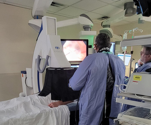
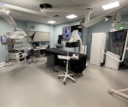
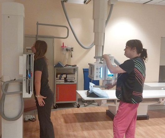
Morton Plant Hospital in Clearwater, FL recently installed a new fluoroscopy system for its endoscopy program. is the most advanced interventional system available and the only ERCP fluoro system proven to reduce radiation exposure by up to 84%. This improves the radiation safety for both staff and patients. The Omega E-View.AI
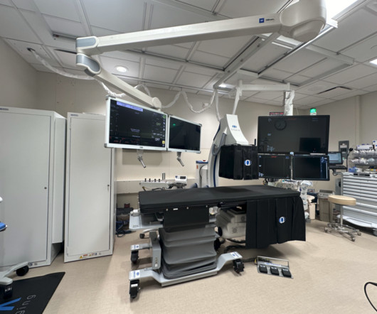
Northwestern Medicine McHenry Hospital in McHenry, IL recently installed a new fluoroscopy system for its endoscopy program. is the most advanced interventional system available and the only ERCP fluoro system proven to reduce radiation exposure by up to 84%. The Omega E-View.AI
Virginia Mason Medical Center in Seattle, WA is installing new fluoroscopy systems for its therapeutic endoscopy program. is an innovative fluoro system proven to reduce radiation exposure by up to 84%. The Omega E-View.AI
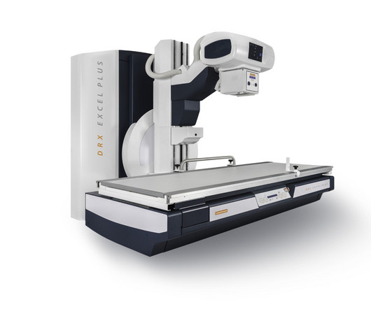
DXR-Excel Plus is a two-in-one system for both fluoroscopy and general radiology that delivers real-time, images for a wide range of exams, while providing features that help create an enhanced experience for users, patients, and administrators, the company said. Image courtesy of Carestream Health.
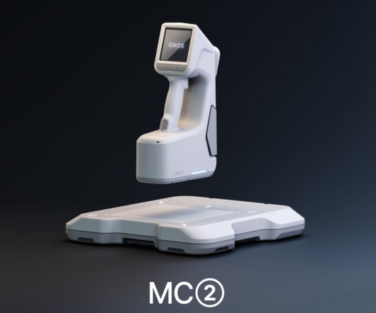
MC2 is cordless and lightweight and offers dynamic digital radiography and fluoroscopy in addition to static imaging capabilities, according to Oxos. It features a patented positioning system designed to assist in alignment for image capture.
Food and Drug Administration (FDA) has said CT is still the preferred imaging modality for patients with medical devices. In an October 15 communication , the agency said it had received reports of electronic medical devices being damaged during CT scans due to radiation. Read the FDA's full guidance here.
Atlanta-based West Physics Consulting said that it has completed the purchase of Radiation Protection Services Ltd., Radiation Protection Services customers will have access to subject matter expertise in the areas of MRI safety, CT and fluoroscopy dose optimization, and clinical image analysis and review for ACR accreditation.
milla1cf Thu, 01/11/2024 - 08:22 January 11, 2024 — Carestream Health has launched a new and enhanced DRX-Excel Plus X-ray System that boosts the performance of the powerful, two-in-one solution to enable more productivity and efficiency, higher image quality, and an improved experience for users and patients.
The evolution of interventional X-ray systems over the past few decades has been remarkable, driven by the pursuit of improving image quality, procedural efficiency, and patient and staff safety.
With the dynamic functional imaging capability of DDR, the authors could visualize thoracic and pulmonary motion and track diaphragm movement. No physician presence is required, radiation dose is lower than an average fluoroscopy exam and the dynamic images can be captured with the patient sitting, standing or on a table.
CT-fluoroscopy (CTF) allows radiologists to acquire near-real-time images with equivalent biopsy accuracy and reduced procedural times, with the tradeoff of patient and physician exposure to low levels of ionizing radiation. Percutaneous CT-guided biopsies are increasingly important in the era of precision medicine.
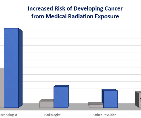
Fluoroscopy-guided procedures are the leading source of occupational ionizing radiation exposure for electrophysiologic (EP) personnel. High cumulative doses of X-ray radiation might increase the risk for malignancies, early development of cataracts and orthopedic problems due to the heavy weight of lead aprons. What they found?
Brigham and Women’s Hospital in Boston recently added radiation-saving technology to their Endoscopy Center. With a full renovation of their two gastroenterology rooms. The E-View.AI The E-View.AI
Continuous-rotation computed tomography (CT) fluoroscopy is an imaging modality widely used in interventional radiology (IR) procedures, facilitating precise punctures even into small lesions and lesions deep within the body by rapid, real-time, and high-resolution tomographic images (1,2).
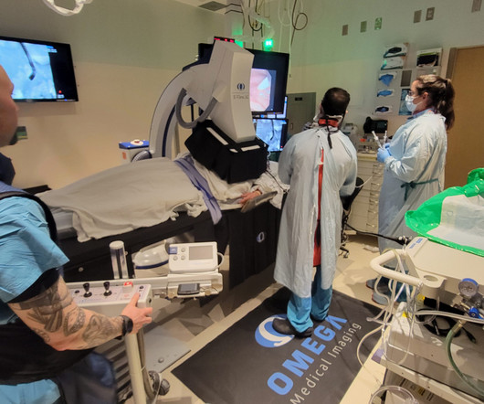
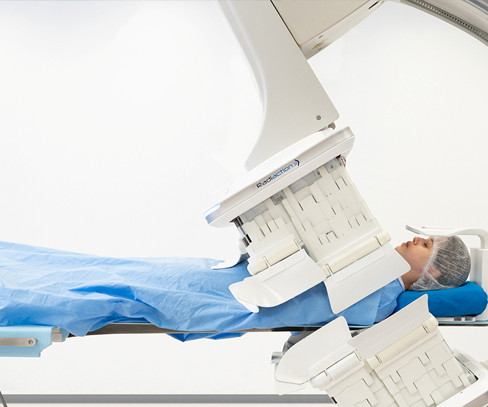
A career in interventional medicine is one that comes with the risks inherent to radiation exposure. Omega Medical Imaging has developed next-level technology that dramatically reduces radiation exposure without compromising image quality or changing existing workflow. But should physicians simply accept that fate?
Here you’ll find a highly skilled, compassionate staff along with comprehensive advanced imaging technologies that are raising the bar for patient care in their rural community. As for magnetic resonance imaging (MRI), just 19% of extremely disadvantaged zip codes had access as compared to 32% of extremely advantaged. with 25 beds.
Spot finding - For spot finding one has to look at a report, form an image and eventually find a differential diagnosis. It is crucial to regularly observe various reports like MRI, CT scan, X-Ray, Doppler, Fluoroscopy etc. Textbook of radiology and imaging- Sutton David ii. Therefore I recommend two methods to approach it- i.
As technology evolved, doctors turned to imaging-guided procedures using computed tomography (CT) or X-ray technology. Considered a minimally invasive procedure, image-guided epidural injections for back pain may be administered at a doctor’s office, surgical center or hospital imaging center.
Radiology is a medical imaging procedure that uses ionizing electromagnetic radiation to create images of bones, organs, and soft tissues to diagnose a patient’s symptoms, disease, or conditions. It includes techniques like X-rays, CT scans, MRIs, ultrasounds, and fluoroscopy.
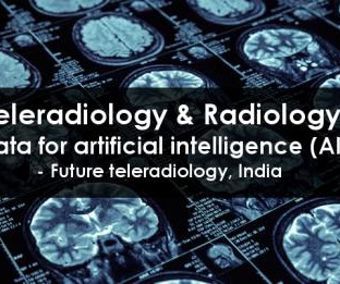
Teleradiology & Radiology data for artificial intelligence (AI) Introduction: “Illuminating Shadows” invites you on a comprehensive journey into the fascinating world of X-ray imaging. Chapter 1: Introduction to X-ray Imaging An overview of the importance of X-ray imaging in healthcare.
Closeup of X-ray photography of human brain Introduction: “The Radiant Thread” is a comprehensive guide that unravels the intricate world of X-ray imaging from A to Z. From its historical roots to contemporary innovations, we will follow the radiant thread that connects all aspects of X-ray imaging.
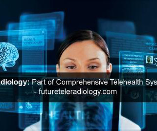
Teleradiology Introduction: “Beneath the Surface” is an illuminating journey that delves deep into the world of X-ray imaging, unveiling its crucial role in patient care. In this exploration, we will dive into the history, technology, and the profound impact of X-ray imaging on healthcare.
This journey takes us from the early days of X-ray discovery by Wilhelm Roentgen to the cutting-edge digital and computational innovations that shape the modern landscape of diagnostic imaging. Chapter 2: The Art and Science of Radiography A closer look at the development of radiography, the first X-ray imaging method.
Introduction to Sports Radiology: A Specialized Discipline: Define sports radiology as a specialized discipline within medical imaging. Discuss the unique challenges and considerations involved in imaging athletes with acute injuries. Arthrography: Contrast-Enhanced Joint Imaging: Explore the use of arthrography in sports radiology.
How X-rays are generated, interact with matter, and produce diagnostic images. Chapter 3: Types of X-ray Technology: Beyond Radiography An exploration of the various modalities and applications of X-ray technology, from radiography to fluoroscopy and computed tomography (CT).
How X-rays are generated, interact with the human body, and create diagnostic images. Chapter 3: The Evolution of Radiography: From Shadows to Images An exploration of the development of radiography, the earliest X-ray imaging technique. How these modalities enhance diagnostic capabilities for specific medical conditions.
Outpatient radiology centers play a crucial role in the healthcare landscape by providing convenient, efficient, and cost-effective access to diagnostic imaging services for patients across a wide range of medical conditions. These services include X-rays, ultrasounds , MRIs, CT scans, mammography, and fluoroscopy, among others.
The following is the list of candidates for the 2024 edition of the Minnies, AuntMinnie.com 's campaign to recognize the best and brightest in medical imaging. Image from Martin W. Image from Denis Le Bihan, PhD, of the NeuroSpin research facility, et al. Image from Richard Carson, PhD, of Yale University, et al.
Standardized guidelines can help radiologists navigate the legal landscape of imaging pregnant women with ionizing radiation, according to a July 15 editorial published in the Journal of the American College of Radiology ( ACR ). The parameter provides best practices for imaging pregnant women or women who can become pregnant.
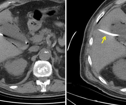
The patient is pregnant (orange arrows), therefore ionizing radiation with CT scan or fluoroscopy can not be used for imaging guidance. The final CT image shows the drainage catheter (yellow arrow) correctly placed in the gallbladder with the tip coiled in the gallbladder fundus (red arrow). MRI of abdomen. J Visc Surg.
We organize all of the trending information in your field so you don't have to. Join 5,000 users and stay up to date on the latest articles your peers are reading.
You know about us, now we want to get to know you!
Let's personalize your content
Let's get even more personalized
We recognize your account from another site in our network, please click 'Send Email' below to continue with verifying your account and setting a password.


Let's personalize your content