This site uses cookies to improve your experience. To help us insure we adhere to various privacy regulations, please select your country/region of residence. If you do not select a country, we will assume you are from the United States. Select your Cookie Settings or view our Privacy Policy and Terms of Use.
Cookie Settings
Cookies and similar technologies are used on this website for proper function of the website, for tracking performance analytics and for marketing purposes. We and some of our third-party providers may use cookie data for various purposes. Please review the cookie settings below and choose your preference.
Used for the proper function of the website
Used for monitoring website traffic and interactions
Cookie Settings
Cookies and similar technologies are used on this website for proper function of the website, for tracking performance analytics and for marketing purposes. We and some of our third-party providers may use cookie data for various purposes. Please review the cookie settings below and choose your preference.
Strictly Necessary: Used for the proper function of the website
Performance/Analytics: Used for monitoring website traffic and interactions
Fluoroscopy-assisted ultrasound guidance for mini-percutaneous nephrolithotomy (mini-PCNL) procedures in children is a safer and more effective approach than fluoroscopy alone, researchers have found. But PCNL does impart radiation, and clinicians have sought to mitigate this exposure, especially to pediatric patients.
A team of researchers at Boston Children’s Hospital has developed an age-specific dose catalog for estimating radiation exposure to children from diagnostic and interventional radiology fluoroscopy procedures. They analyzed metrics to estimate age-specific effective dose per IR procedure type and diagnostic fluoroscopy exam.
Initial percutaneous nephrostomy (PCN) tube placement leads to more radiation exposure for pregnant women with suspected kidney stones, according to a study published October 27 in Urology. Options include ureteral stent, PCN, or primary ureteroscopy, with the potential for multiple subsequent procedures that often use fluoroscopy.
. | M7-SSPH05-2 | Room N229 Findings will be presented in this Monday afternoon presentation on organ-specific ionizing radiation doses in neonatal patients who undergo interventional procedures for congenital heart disease (CHD). Gy-cm2, with organ-specific radiation doses highest for lung from frontal view (8.1 Louis, and colleagues.
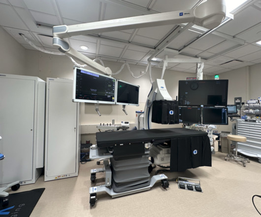
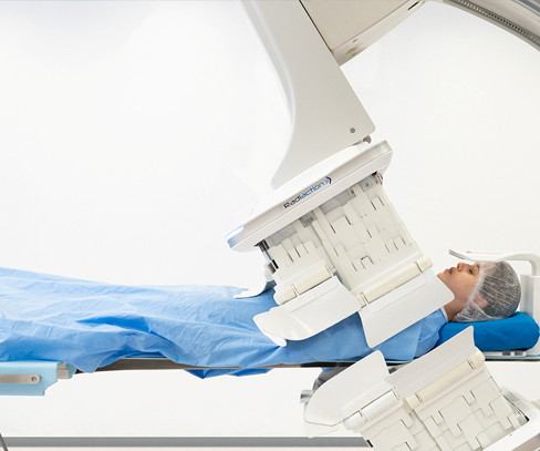
Morton Plant Hospital in Clearwater, FL recently installed a new fluoroscopy system for its endoscopy program. is the most advanced interventional system available and the only ERCP fluoro system proven to reduce radiation exposure by up to 84%. This improves the radiation safety for both staff and patients. The Omega E-View.AI
Northwestern Medicine McHenry Hospital in McHenry, IL recently installed a new fluoroscopy system for its endoscopy program. is the most advanced interventional system available and the only ERCP fluoro system proven to reduce radiation exposure by up to 84%. The Omega E-View.AI
Virginia Mason Medical Center in Seattle, WA is installing new fluoroscopy systems for its therapeutic endoscopy program. is an innovative fluoro system proven to reduce radiation exposure by up to 84%. The Omega E-View.AI
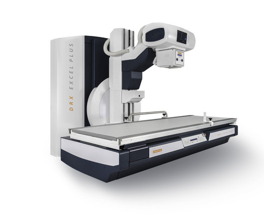
DXR-Excel Plus is a two-in-one system for both fluoroscopy and general radiology that delivers real-time, images for a wide range of exams, while providing features that help create an enhanced experience for users, patients, and administrators, the company said.
Atlanta-based West Physics Consulting said that it has completed the purchase of Radiation Protection Services Ltd., Radiation Protection Services customers will have access to subject matter expertise in the areas of MRI safety, CT and fluoroscopy dose optimization, and clinical image analysis and review for ACR accreditation.
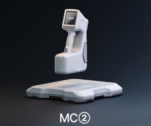
MC2 is cordless and lightweight and offers dynamic digital radiography and fluoroscopy in addition to static imaging capabilities, according to Oxos. MC2s small scatter area and low radiation output can reduce the space and infrastructure needs required by larger systems, the company said.
Read more on AuntMinnie.com Related Reading: Philips issues radiation warning for fluoroscopy, angiography systems Philips begins U.S./European Philips is introducing a new super contrast-enhanced ultrasound (CEUS) applicatio.
In an October 15 communication , the agency said it had received reports of electronic medical devices being damaged during CT scans due to radiation. Interference is when the radiation and the device electronics are incompatible, and the resulting damage causes the device to fail to work normally," the FDA said.
To update normative data on fluoroscopy dose indices in the United States for the first time since the Radiation Doses in Interventional Radiology study in the late 1990s.
Effects of low-dose ionizing radiation on genomic instability in interventional radiology workers. Quantifying Regional Radiation-Induced Lung Injury in Patients Using Hyperpolarized 129Xe Gas Exchange Magnetic Resonance Imaging. Rankine, et al, International Journal of Radiation Oncology, Biology, Physics , September 1, 2023.
CT-fluoroscopy (CTF) allows radiologists to acquire near-real-time images with equivalent biopsy accuracy and reduced procedural times, with the tradeoff of patient and physician exposure to low levels of ionizing radiation. Percutaneous CT-guided biopsies are increasingly important in the era of precision medicine.
Carestream’s DRX-Excel Plus X-ray System is a flexible solution for both fluoroscopy and general radiology that can deliver real-time, high-quality images for a wide range of exams while providing important features that help create an enhanced experience for users, patients, and administrators.
Initially, pulsed fluoroscopy marked a huge shift from continuous fluoroscopy by considerably reducing radiation exposure while preserving diagnostic accuracy.
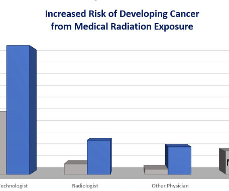
Fluoroscopy-guided procedures are the leading source of occupational ionizing radiation exposure for electrophysiologic (EP) personnel. High cumulative doses of X-ray radiation might increase the risk for malignancies, early development of cataracts and orthopedic problems due to the heavy weight of lead aprons. What they found?
Percutaneous CT fluoroscopy (CTF) guided procedures have been shown to decrease complications and procedural time vs. conventional (out-of-room) CT guidance. However, many physicians forgo the use of in-room CTF due to concerns about operator radiation exposure.
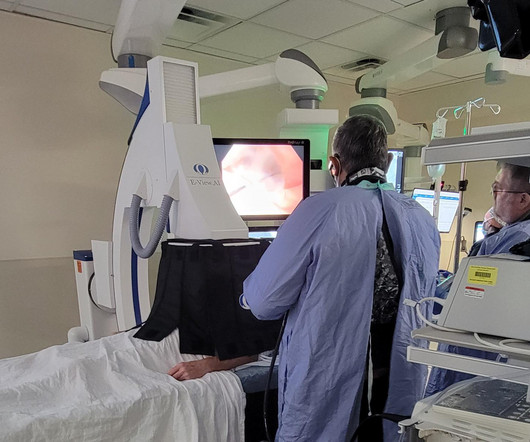
Brigham and Women’s Hospital in Boston recently added radiation-saving technology to their Endoscopy Center. With a full renovation of their two gastroenterology rooms. The E-View.AI The E-View.AI
No physician presence is required, radiation dose is lower than an average fluoroscopy exam and the dynamic images can be captured with the patient sitting, standing or on a table.
Continuous-rotation computed tomography (CT) fluoroscopy is an imaging modality widely used in interventional radiology (IR) procedures, facilitating precise punctures even into small lesions and lesions deep within the body by rapid, real-time, and high-resolution tomographic images (1,2).
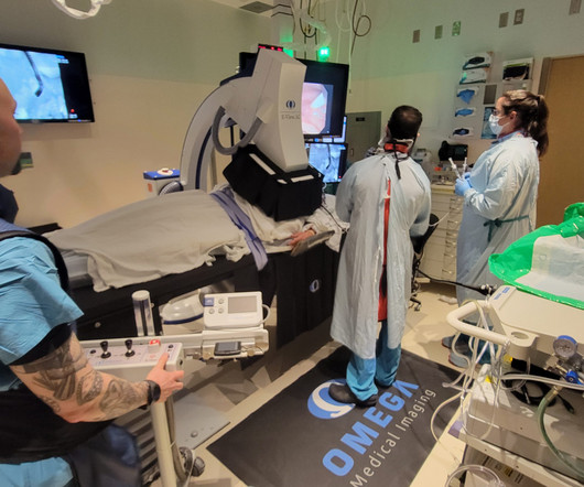
A career in interventional medicine is one that comes with the risks inherent to radiation exposure. Omega Medical Imaging has developed next-level technology that dramatically reduces radiation exposure without compromising image quality or changing existing workflow. But should physicians simply accept that fate?
This procedure, however, involves exposure to ionizing radiation, which raises concerns regarding long-term radiation risks. This study aims to evaluate the radiation exposure and dosimetric outcomes of PAE using n-BCA, focusing on radiation dose-area product (DAP) and fluoroscopy time (FT).
However, concerns regarding radiation exposure remain due to the procedural complexity. This study aims to evaluate the radiation exposure associated with PAE performed in multiple outpatient settings using mobile C-arm fluoroscopy units.
Preliminary results on radiation metrics from this comparative retrospective study on nBCA versus microsphere PAEs are reviewed here to provide data points for further advancement in this realm.
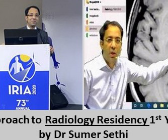
It is crucial to regularly observe various reports like MRI, CT scan, X-Ray, Doppler, Fluoroscopy etc. Also you will learn the operation of various machines which again is not supposed to be your expertise but in exams like the DNB there will be theory papers on radiation physics. It is the skill of keen observation of anomalies.
Radiology is a medical imaging procedure that uses ionizing electromagnetic radiation to create images of bones, organs, and soft tissues to diagnose a patient’s symptoms, disease, or conditions. It includes techniques like X-rays, CT scans, MRIs, ultrasounds, and fluoroscopy.
Patient Radiation Doses in IR Procedures: The American College of Radiology Dose Index Registry-Fluoroscopy Pilot. Corrigendum to Jones AK, et al. J Vasc Interv Radiol 2023; 34:544-555.e11.
The median fluoroscopy time was 22.2 (IQR: minutes, and the median radiation dose was 753 (IQR: 417 - 1559) mGy. This retrospective study evaluates the feasibility, safety, and short-term efficacy of PAE using n-BCA glue embolization in 244 patients from June 2022 through May 2024. IQR: 17.1 – 30.0) weeks for the IPSS (9.5 ± 6.0 [SD]
Role of Imaging In Epidural Injections In the 1980s, radiologists began using fluoroscopy to assist with needle placement. Today, CT or fluoroscopy technology helps guide the needle to the precise injection site. Studies have shown that blind injections may be inaccurate 25 to nearly 40 percent of the time.
How technological advancements have improved image quality, reduced radiation exposure, and expanded diagnostic capabilities. Chapter 5: Beyond Radiography: The Rise of Advanced Modalities An examination of advanced X-ray modalities, including fluoroscopy, mammography, and computed tomography (CT).
The radiologic technologists also like having removable, lightweight detectors because it makes it easier to perform numerous extremity exams and the system also delivers less radiation dose. The portable machine saves a lot of time for us and saves a lot of discomfort for the patient by not having to move them.”
C-Arm Fluoroscopy: Real-Time Imaging in Interventional Procedures: Highlight the use of C-arm fluoroscopy for real-time imaging during interventional procedures. Patient-Centric Imaging: Minimizing Radiation Exposure: Discuss the importance of patient-centric imaging in sports radiology.
The principles of radiation and how X-rays interact with the human body to create diagnostic images. Chapter 3: Types of X-ray Imaging: Beyond Radiography An exploration of the various types of X-ray imaging, including radiography, fluoroscopy, and computed tomography (CT). How each modality is used for different clinical purposes.
Chapter 3: The Radiologic Toolbox – Types of X-ray Imaging An exploration of the various types of X-ray imaging, including radiography, fluoroscopy, computed tomography (CT), and more. The importance of minimizing radiation exposure while maintaining diagnostic accuracy.
Chapter 3: Types of X-ray Technology: Beyond Radiography An exploration of the various modalities and applications of X-ray technology, from radiography to fluoroscopy and computed tomography (CT). The delicate balance between minimizing radiation exposure and achieving precise diagnostic images.
Chapter 4: Beyond Radiography: Advanced X-ray Modalities An examination of advanced X-ray modalities, including fluoroscopy, computed tomography (CT), and mammography. Chapter 6: Radiation Safety: Balancing Benefits and Risks The importance of radiation safety in X-ray technology, including dose management and patient protection.
Chapter 4: Beyond Radiography: Advanced X-ray Modalities An examination of advanced X-ray modalities, including fluoroscopy, computed tomography (CT), and mammography. Chapter 6: Radiation Safety: Balancing Benefit and Risk The importance of radiation safety in X-ray technology, including dose management and patient protection.
These services include X-rays, ultrasounds , MRIs, CT scans, mammography, and fluoroscopy, among others. Regulatory Compliance: Compliance with healthcare regulations and standards, such as those related to patient privacy (HIPAA), radiation safety, and quality assurance, is essential but can be challenging to navigate.
Standardized guidelines can help radiologists navigate the legal landscape of imaging pregnant women with ionizing radiation, according to a July 15 editorial published in the Journal of the American College of Radiology ( ACR ).
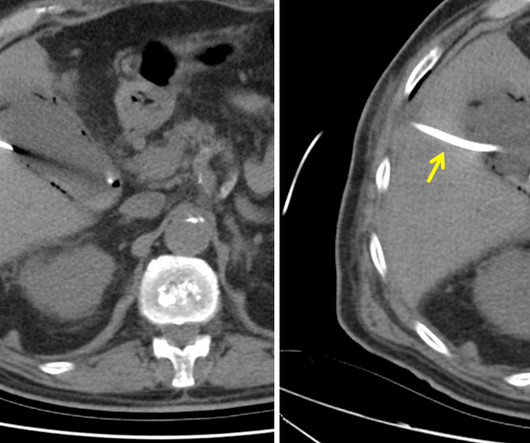
The patient is pregnant (orange arrows), therefore ionizing radiation with CT scan or fluoroscopy can not be used for imaging guidance. What action should be taken for this patient with right upper quadrant pain? MRI of abdomen.
We organize all of the trending information in your field so you don't have to. Join 5,000 users and stay up to date on the latest articles your peers are reading.
You know about us, now we want to get to know you!
Let's personalize your content
Let's get even more personalized
We recognize your account from another site in our network, please click 'Send Email' below to continue with verifying your account and setting a password.


Let's personalize your content