This site uses cookies to improve your experience. To help us insure we adhere to various privacy regulations, please select your country/region of residence. If you do not select a country, we will assume you are from the United States. Select your Cookie Settings or view our Privacy Policy and Terms of Use.
Cookie Settings
Cookies and similar technologies are used on this website for proper function of the website, for tracking performance analytics and for marketing purposes. We and some of our third-party providers may use cookie data for various purposes. Please review the cookie settings below and choose your preference.
Used for the proper function of the website
Used for monitoring website traffic and interactions
Cookie Settings
Cookies and similar technologies are used on this website for proper function of the website, for tracking performance analytics and for marketing purposes. We and some of our third-party providers may use cookie data for various purposes. Please review the cookie settings below and choose your preference.
Strictly Necessary: Used for the proper function of the website
Performance/Analytics: Used for monitoring website traffic and interactions
Fluoroscopy-assisted ultrasound guidance for mini-percutaneous nephrolithotomy (mini-PCNL) procedures in children is a safer and more effective approach than fluoroscopy alone, researchers have found. But PCNL does impart radiation, and clinicians have sought to mitigate this exposure, especially to pediatric patients.
A team of researchers at Boston Children’s Hospital has developed an age-specific dose catalog for estimating radiation exposure to children from diagnostic and interventional radiologyfluoroscopy procedures. They analyzed metrics to estimate age-specific effective dose per IR procedure type and diagnostic fluoroscopy exam.
Initial percutaneous nephrostomy (PCN) tube placement leads to more radiation exposure for pregnant women with suspected kidney stones, according to a study published October 27 in Urology. Options include ureteral stent, PCN, or primary ureteroscopy, with the potential for multiple subsequent procedures that often use fluoroscopy.
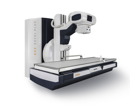
DXR-Excel Plus is a two-in-one system for both fluoroscopy and general radiology that delivers real-time, images for a wide range of exams, while providing features that help create an enhanced experience for users, patients, and administrators, the company said.
To update normative data on fluoroscopy dose indices in the United States for the first time since the Radiation Doses in Interventional Radiology study in the late 1990s.
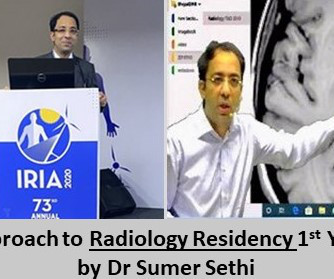
Radiology is a discipline which provides multi-faceted exposure to its pupils. Several post graduates all around the world opt for radiology for this. But it is often confusing for freshers in their initial stage of radiology residency. As far as PG is concerned, the most common practice is to teach Diagnostic radiology.
Carestream’s DRX-Excel Plus X-ray System is a flexible solution for both fluoroscopy and general radiology that can deliver real-time, high-quality images for a wide range of exams while providing important features that help create an enhanced experience for users, patients, and administrators.
12, 2025 Konica Minolta Healthcare Americas, has published a case study by clinicians in the pulmonary and radiology departments at ASST Fatebenefratelli Sacco (Milan, Italy) demonstrating the use of Dynamic Digital Radiography (DDR) to help definitively diagnose diaphragm dysfunction. tim.hodson Fri, 02/14/2025 - 15:14 Feb.12,
Continuous-rotation computed tomography (CT) fluoroscopy is an imaging modality widely used in interventional radiology (IR) procedures, facilitating precise punctures even into small lesions and lesions deep within the body by rapid, real-time, and high-resolution tomographic images (1,2).
Radiology is a medical imaging procedure that uses ionizing electromagnetic radiation to create images of bones, organs, and soft tissues to diagnose a patient’s symptoms, disease, or conditions. It includes techniques like X-rays, CT scans, MRIs, ultrasounds, and fluoroscopy.
Outpatient radiology centers play a crucial role in the healthcare landscape by providing convenient, efficient, and cost-effective access to diagnostic imaging services for patients across a wide range of medical conditions. These services include X-rays, ultrasounds , MRIs, CT scans, mammography, and fluoroscopy, among others.
Radiology plays a pivotal role in diagnosing and managing these injuries, providing valuable insights that guide treatment strategies and facilitate the prompt return of athletes to their respective fields. This blog explores the critical role of radiology in on-the-field diagnostics for sports-related injuries.
Patient Radiation Doses in IR Procedures: The American College of Radiology Dose Index Registry-Fluoroscopy Pilot. Corrigendum to Jones AK, et al. J Vasc Interv Radiol 2023; 34:544-555.e11.
Role of Imaging In Epidural Injections In the 1980s, radiologists began using fluoroscopy to assist with needle placement. Today, CT or fluoroscopy technology helps guide the needle to the precise injection site. Contact Midstate Radiology Associates to schedule an appointment today.
The radiologic technologists also like having removable, lightweight detectors because it makes it easier to perform numerous extremity exams and the system also delivers less radiation dose. “With this machine, the detail is great,” said Frederich Park, MD, Radiologist. What I’m seeing, I see a lot better and with better detail.”
Chapter 2: Basics of X-ray Science – A Radiologic Primer A detailed explanation of the scientific principles that govern X-ray imaging. Chapter 3: The Radiologic Toolbox – Types of X-ray Imaging An exploration of the various types of X-ray imaging, including radiography, fluoroscopy, computed tomography (CT), and more.
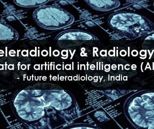
Teleradiology & Radiology data for artificial intelligence (AI) Introduction: “Illuminating Shadows” invites you on a comprehensive journey into the fascinating world of X-ray imaging. The principles of radiation and how X-rays interact with the human body to create diagnostic images.
Chapter 3: Types of X-ray Technology: Beyond Radiography An exploration of the various modalities and applications of X-ray technology, from radiography to fluoroscopy and computed tomography (CT). The delicate balance between minimizing radiation exposure and achieving precise diagnostic images.
How technological advancements have improved image quality, reduced radiation exposure, and expanded diagnostic capabilities. Chapter 5: Beyond Radiography: The Rise of Advanced Modalities An examination of advanced X-ray modalities, including fluoroscopy, mammography, and computed tomography (CT).
Chapter 4: Beyond Radiography: Advanced X-ray Modalities An examination of advanced X-ray modalities, including fluoroscopy, computed tomography (CT), and mammography. Chapter 6: Radiation Safety: Balancing Benefits and Risks The importance of radiation safety in X-ray technology, including dose management and patient protection.
Chapter 4: Beyond Radiography: Advanced X-ray Modalities An examination of advanced X-ray modalities, including fluoroscopy, computed tomography (CT), and mammography. Chapter 6: Radiation Safety: Balancing Benefit and Risk The importance of radiation safety in X-ray technology, including dose management and patient protection.
This year, hundreds of candidates have been selected as semifinalists for 14 categories, ranging from Most Influential Radiology Researcher to Best New Radiology Software. The semifinalist list was compiled based on nominations from members of AuntMinnie.com. Winners will be selected by our expert panel in two rounds of voting.
Standardized guidelines can help radiologists navigate the legal landscape of imaging pregnant women with ionizing radiation, according to a July 15 editorial published in the Journal of the American College of Radiology ( ACR ). The parameter provides best practices for imaging pregnant women or women who can become pregnant.
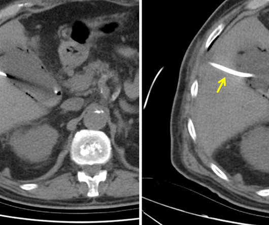
The patient is pregnant (orange arrows), therefore ionizing radiation with CT scan or fluoroscopy can not be used for imaging guidance. He serves as chair for his schools radiology interest group. Rice, MD is the president of Global Radiology CME and is a radiologist with Cape Radiology Group. MRI of abdomen.
We organize all of the trending information in your field so you don't have to. Join 5,000 users and stay up to date on the latest articles your peers are reading.
You know about us, now we want to get to know you!
Let's personalize your content
Let's get even more personalized
We recognize your account from another site in our network, please click 'Send Email' below to continue with verifying your account and setting a password.


Let's personalize your content