This site uses cookies to improve your experience. To help us insure we adhere to various privacy regulations, please select your country/region of residence. If you do not select a country, we will assume you are from the United States. Select your Cookie Settings or view our Privacy Policy and Terms of Use.
Cookie Settings
Cookies and similar technologies are used on this website for proper function of the website, for tracking performance analytics and for marketing purposes. We and some of our third-party providers may use cookie data for various purposes. Please review the cookie settings below and choose your preference.
Used for the proper function of the website
Used for monitoring website traffic and interactions
Cookie Settings
Cookies and similar technologies are used on this website for proper function of the website, for tracking performance analytics and for marketing purposes. We and some of our third-party providers may use cookie data for various purposes. Please review the cookie settings below and choose your preference.
Strictly Necessary: Used for the proper function of the website
Performance/Analytics: Used for monitoring website traffic and interactions
Fluoroscopy-assisted ultrasound guidance for mini-percutaneous nephrolithotomy (mini-PCNL) procedures in children is a safer and more effective approach than fluoroscopy alone, researchers have found. But PCNL does impart radiation, and clinicians have sought to mitigate this exposure, especially to pediatric patients.
Initial percutaneous nephrostomy (PCN) tube placement leads to more radiation exposure for pregnant women with suspected kidney stones, according to a study published October 27 in Urology. Options include ureteral stent, PCN, or primary ureteroscopy, with the potential for multiple subsequent procedures that often use fluoroscopy.
Philips is introducing a new super contrast-enhanced ultrasound (CEUS) applicatio. Read more on AuntMinnie.com Related Reading: Philips issues radiation warning for fluoroscopy, angiography systems Philips begins U.S./European
While conventional chest Xrays and ultrasound can offer clues, additional imaging and tests are often needed for a precise diagnosis, says Michaela Cellina, Head of Imaging Research and a radiologist with ASST Fatebenefratelli Sacco. Diagnosing diaphragm dysfunction is challenging due to its varied symptoms and causes.
It is crucial to regularly observe various reports like MRI, CT scan, X-Ray, Doppler, Fluoroscopy etc. Also you will learn the operation of various machines which again is not supposed to be your expertise but in exams like the DNB there will be theory papers on radiation physics. It is the skill of keen observation of anomalies.
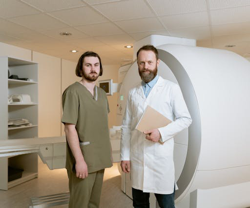
Radiology is a medical imaging procedure that uses ionizing electromagnetic radiation to create images of bones, organs, and soft tissues to diagnose a patient’s symptoms, disease, or conditions. It includes techniques like X-rays, CT scans, MRIs, ultrasounds, and fluoroscopy.
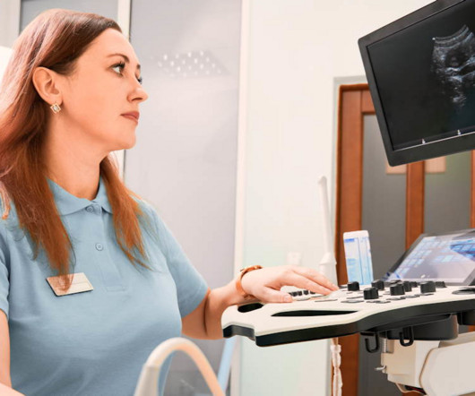
Ultrasound Imaging for Soft Tissue Injuries: Real-Time Visualization: Explore the role of ultrasound in imaging soft tissue injuries. Discuss how real-time visualization with ultrasound aids in assessing muscle, tendon, and ligament injuries, providing immediate feedback.
These services include X-rays, ultrasounds , MRIs, CT scans, mammography, and fluoroscopy, among others. Regulatory Compliance: Compliance with healthcare regulations and standards, such as those related to patient privacy (HIPAA), radiation safety, and quality assurance, is essential but can be challenging to navigate.
Ultrasound model predicts liver disease progression. Appropriateness and imaging outcomes of ultrasound, CT, and MR in the emergency department: a retrospective analysis from an urban academic center. Effects of low-dose ionizing radiation on genomic instability in interventional radiology workers.
Standardized guidelines can help radiologists navigate the legal landscape of imaging pregnant women with ionizing radiation, according to a July 15 editorial published in the Journal of the American College of Radiology ( ACR ).
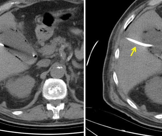
The patient is pregnant (orange arrows), therefore ionizing radiation with CT scan or fluoroscopy can not be used for imaging guidance. Ultrasound of gallbladder used for guidance of percutaneous needle (red arrow) placement for cholecystostomy. Ultrasound of gallbladder demonstrating drainage catheter in the lumen (blue arrow).
We organize all of the trending information in your field so you don't have to. Join 5,000 users and stay up to date on the latest articles your peers are reading.
You know about us, now we want to get to know you!
Let's personalize your content
Let's get even more personalized
We recognize your account from another site in our network, please click 'Send Email' below to continue with verifying your account and setting a password.


Let's personalize your content